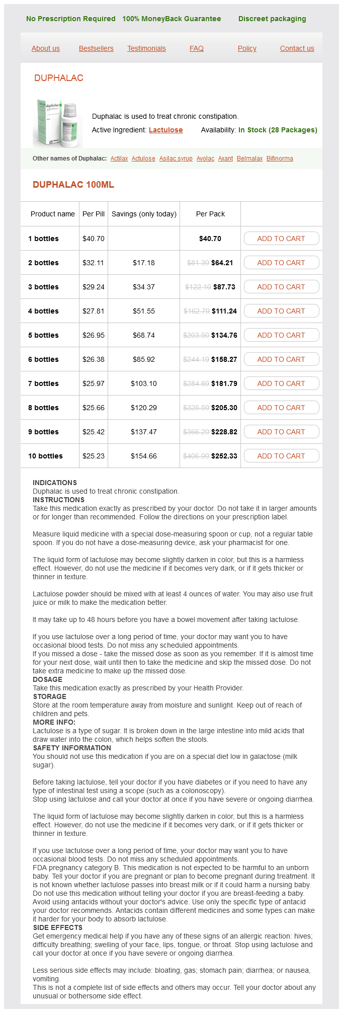
Duphalac
| Contato
Página Inicial

"100 ml duphalac purchase free shipping, treatment interstitial cystitis".
F. Rendell, M.A., M.D., M.P.H.
Clinical Director, Baylor College of Medicine
Following a laminectomy treatment 4 toilet infection order 100 ml duphalac amex, the spinal wire at the thoracic level could additionally be laterally hemisected symptoms ruptured ovarian cyst duphalac 100 ml discount with visa, contused medicine side effects buy cheap duphalac 100 ml. Following lateral hemisection of the spinal wire at level T13 medicine quotes purchase 100 ml duphalac with visa, rats develop bilateral hind paw hypersensitivity to warmth (decreased withdrawal latency) and mechanical (decreased withdrawal threshold) stimuli starting 10 days ater injury. In addition, a bilateral forepaw (above-level) hypersensitivity is observed beginning about 2 weeks ater damage. A few clinically relevant analgesic drugs have been tested in these rats, along with novel remedies. Each rat model displays distinct injury-induced, pain-related behaviors and histopathology-behaviors and pathologies that may not be observed within the different models. Hemisection of the spinal twine might be helpful in evaluating neurochemical and anatomic modifications rostral or caudal to the lesion with the unhurt side. Most spinal wire injuries are as a outcome of acute extradural impact and compressive forces. Following a "reasonable" contusion of the thoracic dorsal spinal twine, the forepaws develop hypersensitivity to warmth and mechanical stimuli, but the sensitivity of the hind paws to these stimuli is variable. Compression Injury To mimic circumferential compression, a mix of impact and compression at multiple ranges somewhat than a single level of the spinal cord, a modiied aneurysm clip is used by which one jaw is slid beneath the anterior thoracic spinal cord, the opposite jaw is over the posterior twine, and the clip is closed for a given length of time. A modiied model of the circumferential compression mannequin was used to evaluate a number of medical analgesic drugs. As with other models, signiicant hind paw hypersensitivity to thermal and mechanical stimuli developed and hind limb locomotor operate diminished; these efects had been noted as early as 1 week ater damage. Behavioral characterization and efect of scientific drugs mannequin of ache following spinal cord compression. Further drug eicacy comparisons between fashions are necessary to help this contention. Ater intravenous injection of erythrosin B, laser irradiation of the thoracic vertebrae causes coagulation of the spinal blood vessels, which is restricted to instant spinal segments. A short-term (several days) mechanical hypersensitivity develops inside the thoracic dermatome during which the ischemia occurred. Furthermore, some of these animals (40%) mutilate their hind limbs ("autotomy"), suggesting the presence of below-level spontaneous ache or dysesthesia. Pharmacologic administration of the phosphodiesterase inhibitor propentofylline, the macrophage migration inhibitory issue inhibitor ibudilast, and the Toll-like receptor 4 antagonist (+)-naltrexone each reversed below-level allodynia bilaterally in this mannequin. On the basis of restricted analyses of spinal lesions utilizing magnetic resonance imaging, a signiicant distinction between patients with ache and pain-free patients and the extent of tract or gray matter harm was not observed. A scientific time course research may not be technically feasible to tackle this concern, nevertheless. At the cellular level, adjustments in excitatory or inhibitory neurotransmission may underlie irregular exercise of, for instance, spinal neurons. However, the indirect pharmacologic proof such as analgesic efects with baclofen suggests that a loss of inhibition might be as necessary as the rise in excitation. Noninvasive magnetic resonance spectroscopy might be used to evaluate modifications in inhibitory amino acids. Spinal wire tissue taken acutely following injury (15 to 60 days) exhibited marked microglia/ macrophage iniltration proximal and distal to the damage. Gliopathy has been deined as the "dysfunctional and maladaptive" glia response to injury. Chapter 107 Chronic Pain: Basic Science 1951 Gliopathy has also been reported in different neuropathic ache models. Glial Inhibitors A variety of intracellular or membrane targets modulate the glial response to injury. Postmitotic cells such as oligodendrocytes and neurons reply to these proliferation alerts by present process apoptosis. One such class of lipids is the lysophospholipids, lipids involved in cell proliferation and chemotaxis. Drugs with multiple mechanisms might subsequently be fascinating over those with a speciic mechanism. A key check of the usefulness of a preclinical mannequin is whether or not or not it can predict the clinical eicacy of a given treatment. Researchers consider that their models may be generalized to a given medical state of affairs, but this will result in clinical failure on the premise of incorrect assumptions. On the basis of a evaluate of the preclinical and scientific literature, such conidence is lacking. The beneicial efects of glial inhibition need to be timed and balanced to ameliorate pain-a full ablation of glial response has related outcomes as an unresolved glial response. The presence of abnormal evoked or spontaneous neural activity was used to information dorsal root entry zone lesioning. Abnormal single-unit activity recorded in the somatosensory thalamus of a quadriplegic affected person with central ache. Hospital, pharmacy, and outpatient prices for osteoarthritis and persistent again ache. A longitudinal examine of the prevalence and traits of ache within the irst 5 years following spinal wire harm. Pain report and the relationship of ache to bodily elements within the irst 6 months following spinal twine damage. Visceral pain and life high quality in persons with spinal wire harm: a brief report. Chronic pain and nonpainful sensations ater spinal cord injury: is there a relation Neuropathic ache: are there distinct subtypes relying on the aetiology or anatomical lesion Ethnic diferences in pain perception and patient-controlled analgesia utilization for postoperative pain. Evidence-based evaluate of the literature on intrathecal delivery of ache treatment. Behavioral and autonomic correlates of the tactile evoked allodynia produced by spinal glycine inhibition: efects of modulatory receptor techniques and excitatory amino acid antagonists. Inlammatory ache hypersensitivity mediated by phenotypic change in myelinated primary sensory neurons. Chronic peripheral nerve part ends in a rearrangement of the central axonal arborizations of axotomized A beta primary aferent neurons in the rat spinal twine. Subarachnoid transplant of a human neuronal cell line attenuates persistent allodynia and hyperalgesia ater excitotoxic spinal twine damage within the rat. Prediferentiated embryonic stem cells forestall persistent ache behaviors and restore sensory perform following spinal twine damage in mice. Maintenance of train participation in individuals with spinal wire injury: efects on high quality of life, stress and ache. An intensive locomotor coaching paradigm improves neuropathic ache following spinal wire compression injury in rats. Controlling neuropathic pain by adeno-associated virus pushed manufacturing of the anti-inlammatory cytokine, interleukin-10. Histogranin, a modiied histone H4 fragment endowed with N-methyl-D-aspartate antagonist and immunostimulatory activities. Clinical feasibility for cell therapy utilizing human neuronal cell line to treat neuropathic behavioral hypersensitivity following spinal twine damage in rats. Biomaterial-based interventions for neuronal regeneration and functional restoration in rodent model of spinal wire injury: a scientific evaluation. Combination of engineered Schwann cell grats to secrete neurotrophin and chondroitinase promotes axonal regeneration and locomotion ater spinal twine injury. Strategies for regeneration of components of nervous system: scafolds, cells and biomolecules. Cell transplantation and neuroengineering strategy for spinal wire injury remedy: a summary of present laboratory indings and evaluation of literature. Efects of adrenal medullary transplants on pain-related behaviors following excitotoxic spinal wire harm. Serotonergic neural precursor cell grats attenuate bilateral hyperexcitability of dorsal horn neurons ater spinal hemisection in rat. Sustained analgesic peptide secretion and cell labeling utilizing a novel genetic modiication. Viral vectors encoding endomorphins and serine histogranin attenuate neuropathic ache symptoms ater spinal twine damage in rats.
Diseases
- Right atrium familial dilatation
- Single ventricular heart
- Pancreatic islet cell neoplasm
- Ectrodactyly ectrodermal dysplasia
- Aromatase deficiency
- Enolase deficiency type 3

Several studies have reported recompression on the web site of the vertebroplasty/kyphoplasty medications during pregnancy chart duphalac 100 ml purchase on line. Technical factors treatment wrist tendonitis order 100 ml duphalac, corresponding to poor contact of the cement with the endplate treatment 1st degree heart block buy duphalac 100 ml line, can also be a factor treatment jokes order 100 ml duphalac. By preserving movement, biomechanical stresses at adjacent ranges are closer to an unaltered spine than a fused backbone. Noninferiority research have shown that lumbar and cervical disc replacements provide equal outcomes to traditional fusion strategies for ache reduction and improved perform. Interspinous Devices Interspinous stabilization units have been developed as nonfusion options to augment decompression surgery within the lumbar spine. One device has been associated with a signiicantly larger reoperation rate (33%) compared to minimally invasive decompression and poor cost-efectiveness primarily as a end result of the value of the implant. Pedicle screws are inserted above and under a motion section, then linked by a nonelastic band. Long-term follow-up means that hyperextension and stabilization with the Graf gadget might narrow the spinal canal and exit foramina. When a cavity is created by inlating a balloon in the vertebral physique and subsequently illed with bone cement, the process known as a kyphoplasty. Lateral Interbody Fusion Adjacent-level spinal stenosis is brought on by facet and ligamentum lavum hypertrophy, buckling of the ligament, lack of disc peak, foraminal stenosis, instability, and disc pathology. In a research of 21 patients with stenosis on the rostral level to a fusion, again and leg ache scores were signiicantly decreased. Blood loss is minimal and open decompression of a scarred dural sac could be avoided. Cortical Bone Trajectory Screws to Add on to a Fusion Adding on to a preexisting fusion might require a protracted publicity, in depth dissection through scar tissue, and reinstrumentation of the previous and new spinal segments. A cortical bone trajectory screw is added to the pedicle with a preexisting conventional pedicle screw. If profitable, the technique requires solely restricted posterior publicity and permits an intensive decompression of the adjacent level stenosis. Proliferation of spinal fusions over the last decade has prompted much investigation into the causes, mitigating elements, and therapy of oten very complex spinal issues. Revision surgical procedure for adjacent-level illness occurs at an annual price of about 2-3% per yr. Revision surgical procedure above a fusion can encompass laminectomy alone or laminectomy plus extension of the fusion. Incidence and prevalence of surgical procedure at segments adjacent to a previous posterior lumbar arthrodesis. Incidence, mode and placement of acute proximal junctional failures after surgical treatment of adult spinal deformity. Acute proximal junctional failure in sufferers with preoperative sagittal imbalance. Comparison of adjoining section disease after minimally invasive or open transforaminal lumbar inter body fusion. Minimally invasive lateral interbody fusion for the therapy of rostral adjacent segment lumbar degenerative stenosis without supplemental pedicle screw ixation. What is the rate of lumbar adjacent segment illness after percutaneous versus open fusion Identiication of choice criteria for revision surgical procedure among patients with proximal junctional failure after surgical treatment of spinal deformity. Seven to 10 12 months end result of decompressive surgery for degenerative lumbar spinal stenosis. Adjacent segment illness within the lumbar spine following diferent therapy interventions. Adjacent stage intradiscal strain and segmental kinematics following a cervicl whole disc arthroplasty: an in vitro human cadaveric model. Degeneration and mechanics of the segment adjacent to a lumbar spine fusion: A biomechanical analysis (doctoral dissertation). Biomechanical adjustments at adjacent segments following anterior lumbar inter body fusion using tapered cages. Correlation between sagittal airplane modifications and adjoining phase degeneration following lumbar spine fusion. Spinal stenosis reoperation rate in Sweden is 11% at 10 years-a national analysis of 9664 operations. Relation between laminectomy and development of adjacent segment instability ater lumbar fusion with pedicle ixation. Study on efect of graded facetectomy on change in lumbar movement segment torsional lexibility using three-dimensional continuum contact representation for aspect joints. Lumbar motion segment pathology adjacent to thoracolumbar, lumbar and lumbosacral fusions. Recollapse of previous vertebral compression fracture ater percutaneous vertebroplasty. Recompression of vertebral bodies ater balloon kyphoplasty for vertebral compression fractures- preliminary report. Predictive factor for proximal junctional kyphosis in lengthy fusions to the sacrum in adult spinal deformity. Incidence, threat components, and natural course of proximal junctional kyphosis: surgical outcomes evaluation of grownup idiopathic scoliosis. Five 12 months adjacent stage degeneration modifications with single level disease handled using lumbar total disc replacement with ProDisc-L versus circumferential fusion. Lumbar adjoining section degeneration and disease ater arthrodesis and whole disc arthroplasty. Comparing cost-efectiveness of X-Stop with minimally invasive decompression in lumbar spinal stenosis: a randomized controlled trial. X-Stop versus decompressive surgery for lumbar neurogenic intermittent claudication: randomized managed trial with 2-year follow-up. Does a interspinous device (Colex) improve the outcome of decompressive surgical procedure in lumbar spinal stenosis Rational, rules and experimental evaluation of the concept of sot stabilization. Dynamic stabilization in the surgical administration of painful lumbar spinal issues. Radiographic outcomes of adult spinal deformity correction: a important evaluation of variability and failures across deformity patterns. Instrumentation-related complications of multilevel fusions for grownup spinal deformity patients over age sixty five:surgical considerations and remedy options in sufferers with poor bone high quality. Identiication of determination criteria for revision surgery among sufferers with proximal junctional failure ater surgical remedy of spinal deformity. Incidence, mode and location of acute proximal junctional failures ater surgical therapy of adult spinal deformity. Proximal junctional kyphosis in grownup spinal deformity following lengthy instrumented posterior spinal fusion: incidence, outcomes, and risk issue evaluation. Balloon kyphoplasty and vertebroplasty for Vertebral Compression fractures: a comparative systematic evaluation of eicacy and security. Comparison of vertebroplasty and balloon kyphoplasty for therapy of vertebral compression fractures: a met-analysis of the literature. Balloon kyphoplasty outcomes group balloon kyphoplasty for symptomatic vertebral physique compression fractures ends in speedy, signiicant, and sustained enhancements in again ache, operate, and quality of life for aged patients. Primary and secondary osteoporosis incidence of subsequent vertebral compression fractures ater kyphoplasty. Risk factors of creating new symptomatic vertebral compression fractures ater percutaneous vertebroplasty in osteoporotic sufferers. Subsequent vertebral fractures ater vertebroplasty: affiliation with intraosseous clets. Nitinol spring rod dynamic stabilization system and Nitinol memory loops in surgical treatment for lumbar disc issues: short-term follow-up. Clinical expertise with the Dynesis semirigid ixation system for the lumbar spine: surgical and patient-oriented outcome in 50 circumstances ater a median of 2-years. Posterior pedicle ixation-based dynamic stabilization units for the treatment of degenerative diseases of the lumbar backbone. Adjacent phase degeneration ater lumbar dynamic stabilization utilizing pedicle screws and a nitinol spring rod system with 2-year minimum follow-up. Motion-preserving surgical procedure can prevent early breakdown of adjoining segments: comparison of posterior dynamic stabilization with spinal fusion.

Correction of iatrogenic cervical kyphosis within the presence of a circumferential fusion could additionally be carried out with an anteroposterior procedure with using an appropriately sized and shaped anterior graft and a plate that permits screw translation and angulation ad medicine buy duphalac 100 ml overnight delivery. Of-center placement of anterior cervical distraction pins symptoms 0f low sodium 100 ml duphalac generic, if not acknowledged treatment synonym 100 ml duphalac trusted, could create iatrogenic cervical malalignment within the coronal aircraft medicine in the 1800s purchase duphalac 100 ml line. Aggressive decompressive laminectomies with out fusion, particularly in youngsters, may lead to iatrogenic cervical kyphosis. A corpectomy at the similar level as a previous laminectomy creates signiicant instability; circumferential ixation is normally essential on this state of affairs. Caspar pin misplacement could result in angular, rotational, or translational deformities. Anterior cervical grafts should be massive sufficient to permit for 1 or 2 mm of subsidence with out creating kyphosis. Correction of a cervical scoliosis, with out appreciation for a extreme thoracic curve, may result in the top being tilted to one aspect. Traumatic cervical backbone accidents ixed with inadequate instrumentation or bony buy may result in postoperative malalignment. The signiicant instability that this introduces mandates circumferential fusion in addition to sufficient postoperative immobilization. When circumferential therapy of iatrogenic cervical deformities is required, we generally favor going in anteriorly, decompressing, and putting in undersized grafts and buttress plates to permit extra correction posteriorly, then stepping into posteriorly to further correct and lock within the construct. Correction of cervical scoliosis is associated with a high incidence of root palsies on the concave side. This is probably because of a stretch of the foundation or plexus and appears to recover over a period of weeks. Increased rate of arthrodesis with strut grafting after multilevel anterior cervical decompression. This paper reports a higher rate of fusion amongst sufferers who underwent corpectomy with strut grafting compared with patients treated with multiple cervical discectomies and interbody grafts. The authors report an elevated price of fusion after anterior cervical plating and discectomy with allogenic interbody graft with using a plate. Development of adjacent-level ossiication in patients with an anterior cervical plate. Increased fee of arthrodesis with strut grating ater multilevel anterior cervical decompression. Comparison of Smith-Petersen osteotomy versus pedicle subtraction osteotomy versus anterior-posterior osteotomy types for the correction of cervical spine deformities. The authors report a excessive fee of complications after corpectomy in sufferers who had a prior laminectomy and conclude that circumferential ixation could also be essential in these sufferers. This paper describes the importance of limiting the extent of facetectomy to protect segmental stability in the cervical spine. A standardized nomenclature for cervical backbone sot-tissue release and osteotomy for deformity correction: scientific article. Cobb methodology or Harrison posterior tangent methodology: which to select for lateral cervical radiographic analysis. Anterior cervical plating enhances arthrodesis ater discectomy and fusion with cortical allograt. Furthermore, on the premise of speciic radiographic or pathologic indings, arachnoiditis can be termed arachnoiditis ossiicans, calciic arachnoiditis, or pachymeningitis. In 1951, Smolik and Nash5 recognized that when the outer arachnoid layer is injured, each the blood vessels and mesenchymal cells lend themselves to intensive proliferation. When the arachnoid membrane is uncovered to an insult, an inlammatory response ensues, characterized by ibrinous exudates, neovascularization, and a relative paucity of inlammatory mobile exudates. During this stage, nerve root swelling decreases and the nerve roots adhere to one another and to the pia-arachnoid. Yamagami and colleagues13 postulated that the pathologic modifications in arachnoiditis could also be secondary to diminished dietary provide. However, despite sufficient therapy of the causative agent, scarring of the arachnoid membrane may lead to everlasting harm. Digiovanni and colleagues20 described that the position of an autologous blood patch into the epidural area produced no more inlammation than a standard lumbar puncture. Other authors, although, have described cases by which an epidural blood patch had allegedly been liable for arachnoiditis. Iophendylate (Myodil, Pantopaque) is an oil-based contrast medium utilized in diagnostic myelograms. It was irst used in the United States, in 1944, and its utilization continued for forty years. In Sweden, iophendylate was banned from clinical use in 1948 secondary to animal studies that identiied it as a causative agent for arachnoiditis. Intrathecal injection of corticosteroids was previously used for multiple sclerosis. Boiardi and colleagues32 described several instances of arachnoiditis ater administration of bupivacaine with epinephrine. Gemma and colleagues33 described a case of arachnoiditis ater intrathecal administration of bupivacaine with out epinephrine. It is unclear in these instances whether the arachnoiditis was triggered by the bupivacaine or different preservatives. Smolik and Chapter 106 Arachnoiditis and Epidural Fibrosis 1937 Nash5 showed that easy dural retraction for the visualization of a ruptured intervertebral disc could set off arachnoiditis. Haughton and colleagues35 confirmed that the nucleus pulposus of an intervertebral disc was in a position to cause focal arachnoiditis in monkeys. In obtaining a medical historical past from a patient with arachnoiditis, the clinician should search three main traits of the pain. In many sufferers, arachnoiditis is asymptomatic and is discovered as an incidental radiographic inding. Burton adopted one hundred sufferers with arachnoiditis and located little motor weak point to be present. Guyer and colleagues25 adopted fifty one patients over more than 10 years and located that a decreased vary of movement of the trunk was the most common inding on bodily examination. In cases of continual arachnoiditis with resultant syrinx formation, physical examination indings of syringomyelia are present. Clear proof of arachnoiditis is proven by the thickened, "clumped" nerve roots that no longer present the conventional illing of the nerve root sleeves. Radiographic Features Ater a historical past and physical examination, radiographic imaging research are used to conirm the clinical impression of arachnoiditis. On myelography, two distinct patterns of radiographic arachnoiditis can be diferentiated. In type 2 arachnoiditis, some proliferation is added inside the dural sac, which may be localized or difuse. There are several subtypes of spinal arachnoiditis ossificans based mostly on imaging characteristics. Type 1 has a semicircular arrangement, type 2 is round, and sort 3 demonstrates englobing of the caudal fibers. Spinal epidural ibrosis is caused when ibroblasts from damaged paraspinal muscular tissues enter the vertebral canal and proliferate, forming intensive epidural scarring. Treatment A variety of therapies are aimed at stopping or treating arachnoiditis or epidural ibrosis. Much of the analysis aimed at preventing failed again syndrome has handled strategies to prevent epidural ibrosis. In a rat mannequin of spinal epidural ibrosis, the administration of tissue plasminogen activator helped to prevent postlaminectomy epidural ibrosis. In a canine postlaminectomy mannequin, it has been shown that a single fraction of seven-hundred cGy external beam radiation helped to stop epidural ibrosis and arachnoiditis. Patients were adopted for 1 yr and have been assessed by questionnaires containing the ache scale. Surgical procedures which have been used to treat arachnoiditis embrace spinal fusion procedures, decompressive spinal procedures with out fusion, neuroablative procedures, and implantation of spinal cord stimulators.

A review of the neurological and neurosurgical implications of tuberculosis in youngsters medications that cause weight loss discount duphalac 100 ml fast delivery. Tuberculous lumbar arachnoiditis mimicking conus cauda tumor: a case report and review of literature medicine in french order duphalac 100 ml with mastercard. Cervical intramedullary tuberculoma: acute presentation and rapid response to medical therapy everlast my medicine buy duphalac 100 ml on-line. Large intramedullary abscess of the spinal wire associated with an epidermoid cyst without dermal sinus symptoms for pink eye generic 100 ml duphalac. Pyomyelia: an intramedullary spinal abscess complicating lumbar lipoma with spina biida. Intramedullary spinal cord abscess and subsequent granuloma formation: a rare complication of vertebral osteomyelitis detected by difusion-weighted magnetic resonance imaging. Metachronous occurrence of an intramedullary abscess following radical excision of a cervical intramedullary pilocytic astrocytoma. Multiple nocardial abscesses of cerebrum, cerebellum and spinal wire, inflicting quadriplegia. Pyogenic and non-pyogenic spinal infections: emphasis on difusion-weighted imaging for the detection of abscesses and pus collections. Sequential magnetic resonance imaging inding of intramedullary spinal twine abscess together with 77. Spinal cord problems of tuberculous meningitis; a scientific and pathological examine. Tuberculous meningitis with dementia as the presenting symptom ater intramedullary spinal twine tumor resection. Central nervous system Aspergillus infection ater epidural analgesia: prognosis, therapeutic challenges, and literature review. Magnetic resonance imaging screening to establish spinal and paraspinal infections related to injections of contaminated methylprednisolone acetate. Spinal and paraspinal fungal infections related to contaminated methylprednisolone injections. Syndrome of the anterior spinal artery as the primary manifestation of aspergillosis. Chronic mycotic meningitis with spinal involvement (arachnoiditis): a report of ive circumstances. Rapidly progressive quadriparesis heralding disseminated coccidioidomycosis in an immunocompetent patient. Spinal twine compression because of intradural extramedullary aspergilloma and cyst: a case report. Spinal arachnoiditis due to Aspergillus meningitis in a beforehand wholesome patient. Most spinal vascular lesions are characterized by an irregular arteriovenous shunt, which may be situated inside the dura, on the spinal twine floor, inside the substance of the spinal wire or, not often, extradurally. Whereas the latter lesions are sometimes congenital, the more frequent istulous lesions are oten acquired. A fundamental understanding of the anatomy and pathophysiology of those lesions coupled with reinements in endovascular and operative methods has permitted profitable deinitive therapy with minimal morbidity in most sufferers harboring vascular shunts of the spinal wire. Early surgical therapy was directed at stripping the long dorsal vein of the spinal cord floor. It was assumed that tiny feeding vessels, too small to be seen angiographically, equipped this dilated vein all through its size. As talked about earlier, these shunts and their venous drainage are almost universally dorsally located, with ventral communications being exceedingly rare. Other metameric anomalies of associated organs and the pores and skin are commonly associated with these lesions. Gueguen and colleagues23 in addition to Anson and Spetzler12 subdivided these arteriovenous shunts based on the complexity and measurement of the lesion. Similarly, the presenting signs and signs of spinal vascular malformations depend on whether or not the pertinent pathophysiologic process is venous congestion, hemorrhage, ischemia, or localized mass efect. Each sort of lesion is mentioned individually on this section, however, normally, comparatively slow-low lesions have sometimes chronic progressive courses indicative of excessive venous pressures, whereas high-low lesions oten have more abrupt shows related to hemorrhage or ischemia. Because spinal twine perfusion pressure is equal to imply systemic arterial stress minus venous strain, progressive spinal wire ischemia could ensue. Episodic acute neurologic deterioration in these sufferers may happen on account of venous thrombosis. In uncommon circumstances, sufferers may current with subarachnoid hemorrhage mimicking that of intracranial aneurysm rupture. Symptoms sometimes come up insidiously with again and leg pain and delicate sensorimotor dysfunction. In early levels, symptoms might mimic these of neurogenic claudication secondary to spinal stenosis. Neurologic examination, nonetheless, oten reveals blended higher and lower motor neuron illness and patchy sensory loss, clearly diferentiating the clinical picture from degenerative lumbar stenosis. If untreated, these lesions lead to signiicant incapacity and wheelchair dependence in most patients inside 6 months to 3 years ater symptom onset. However, in more continual circumstances, progressive ischemia ultimately results in irreversible neuronal loss and infarction. For instance, typical headache and nuchal rigidity will be the only signs of a spinal subarachnoid hemorrhage and usually result in the usual evaluation for an intracranial ruptured aneurysm. Symptoms could come up at any age, although most patients current in childhood and early to middle adult years. Associations with hereditary hemorrhagic telangiectasia (Rendu-Osler-Weber disease) and Kartagener syndrome have been noted in younger patients. In addition to such "vascular steal" mechanisms, the excessive stress and low characteristics via these dysmorphic vessels render them susceptible to hemorrhage, typically on the venous aspect of the malformation, oten from a venous aneurysm. Finally, enlarged tortuous feeding vessels or draining veins may cause localized mass efect with compression of the spinal twine or spinal roots, leading to myelopathy or radiculopathy, respectively. An acute presentation from subarachnoid hemorrhage or intramedullary hemorrhage is most typical. If the illing defect extends toward a neural foramen, the level and side of the istula could also be suggested. For advanced instances, intraoperative conirmation of shunt elimination may be obtained. Should the latter not be possible, a dural substitute, both as onlay or sutured grat, might serve to scale back the danger of cerebrospinal luid leak and postoperative scarring from the paraspinal musculature. Advances in catheter expertise, image decision, and embolization materials have allowed increasing success and security within the elimination of sure vascular lesions. Both neurophysiologic monitoring and pharmacologic provocative testing have emerged as essential adjuncts in the endovascular remedy of spinal vascular malformations. Surgical Therapy: General Considerations he surgical strategy to spinal wire vascular malformations is determined by the level and anatomic place of the lesion. Nevertheless, the majority are dorsal or dorsolateral and therefore can be approached by way of a regular posterior laminectomy of applicable variety of levels. In basic, routine perioperative antibiotics and corticosteroids are administered on the time of surgical procedure. Neurophysiologic monitoring, including somatosensory evoked potential and motor evoked potential, are also routinely used. Whereas authors such as Malis53 have described using the sitting or oblique positions for the posterior removing of malformations, we choose the inclined position for all such laminectomies. Although the sitting place decreases venous stress and respiratory excursions, it also precludes the efective use of an assistant during the operation. With the patient within the inclined position, the surgeon and assistant work together throughout the working table. Operative stripping of the long dorsal vein of the spinal twine surface was recognized as a tedious and pointless procedure that was answerable for neurologic morbidity as a outcome of removal of regular spinal twine venous drainage. Currently, these lesions are handled by less complicated and more reined surgical technique or by endovascular occlusion. A paramedian longitudinal dural incision permits exposure of the intradural nerve root and initial phase of the associated draining vein of the shunt. Simple interruption of the draining vein is the widely most popular technique, particularly in circumstances in which the radicular artery that provides the istula also supplies a spinal cord medullary artery. Several millimeters of the feeding radicular artery and intradural draining vein could additionally be cauterized, divided, and contiguously excised along with a small window of dura on the foundation sleeve.
Order 100 ml duphalac free shipping. Do YOU Have a Generalized Anxiety Disorder (GAD)? 6 Signs and Symptoms.