
Dutasteride
| Contato
Página Inicial
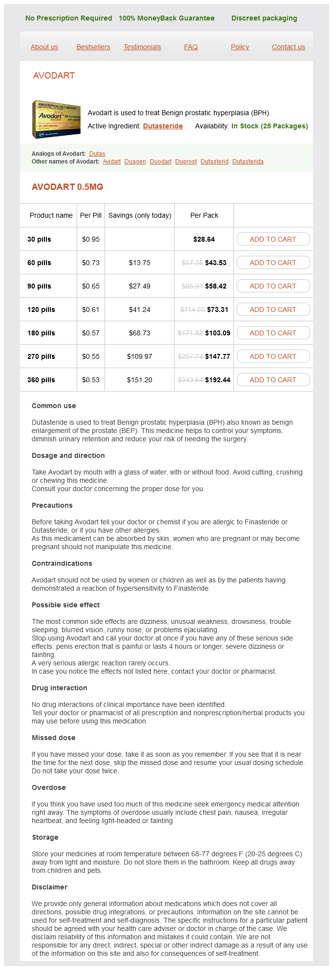
"0.5 mg dutasteride best, hair loss in men zip off pants".
F. Frithjof, M.A., Ph.D.
Co-Director, Idaho College of Osteopathic Medicine
Continue to advance the insertion twine tip via the vocal cords and to a degree approximately 3 cm above the carina hair loss mens health dutasteride 0.5 mg buy cheap. It could be very likely that the insertion wire has entered the esophagus if the 15 cm mark has been passed hair loss cure 3d dutasteride 0.5 mg buy otc. Position the tip of the insertion wire just above the tip of the epiglottis german hair loss cure cheap 0.5 mg dutasteride mastercard, then advance it a quantity of millimeters posterior to the epiglottis whereas angulating the tip of the insertion cord barely anterior by urgent down on the angulation lever hair loss in men engagement generic dutasteride 0.5 mg online. Simultaneously rotate, angulate, and advance the insertion cord tip towards and previous the vocal cords. This approach has the benefit of bringing the tip of the insertion wire instantly midline and towards the epiglottis. This can cause some patient discomfort early within the procedure and should decrease affected person cooperation before the insertion twine has entered the trachea. There is the potential for epistaxis to make visualization of laryngeal constructions difficult if not impossible. It is troublesome to anesthetize the nasal passages fully despite the applying of topical anesthesia to the nasal passages. Visualization of the epiglottis (at the highest of the photo) via the flexible fiberoptic bronchoscope. The tip of the insertion cord could stay angulated and abut against the wall of the trachea. The insertion cord tip could also be abutting the arytenoid cartilages or the pyriform sinus. If inadequate anesthesia is the trigger, inject 2 mL of 2% or 4% lidocaine by way of the working channel of the insertion cord and wait several minutes for it to take effect. Having the patient encourage deeply will deliver the vocal cords into higher opposition. Once the insertion wire tip has passed the vocal cords, bring the tip into impartial position with a light downward motion of the angulation lever. Advance the insertion wire tip further to convey the bifurcation of the trachea at the carina into view. The trachea can easily be identified anteriorly by the cartilaginous rings and posteriorly by the graceful mucosa of the posterior wall. This is achieved by advancing the tip of the insertion wire to the carina with the thumb and forefinger of the nondominant hand. Withdraw the insertion twine till the gap on the insertion cord between the marked level and the tracheal tube connector is four cm. Oral fiberoptic intubation requires that the insertion cord tip traverse a more acute angle to reach the vocal cords than it would by the nasal route. A method believed to considerably enhance the first-time pass rate with oral fiberoptic bronchoscopic intubation is to cross a lubricated 5. This permits an assistant to maintain the face masks and ventilate the patient during the fiberoptic bronchoscopy. Additionally, air flow may be maintained the whole time just by using a bag-valve system linked to the 5. Failure to intubate could be as a end result of narrow nasal or airway passages, blood or vomitus limiting the fiberoptic subject of view, or inexperience. Oxygen insufflation into the pharynx and esophagus works its means into the abdomen. A randomized managed trial confirmed no difference within the incidence of vocal wire damage for nasotracheal fiberoptic intubation versus orotracheal intubation. Intubation via the fiberoptic bronchoscope is an option to contemplate when direct laryngoscopy is tough or impossible. Awake fiberoptic intubation may be preferable in some cases to different methods that render the patient unconscious and apneic. Fiberoptic intubation is related to a high success rate when performed by appropriately trained people. The advantages of performing regional anesthesia of the oropharynx, larynx, and trachea previous to fiberoptic intubation are a quiet visual subject and improved affected person acceptance. In most instances, fiberoptic intubation is a particularly protected and efficient technique of securing the airway. Elizondo E, Navarro F, Perez-Romoa, et al: Endotracheal intubation with versatile fiberoptic bronchoscopy in sufferers with irregular anatomic situations of the head and neck. Dabbagh A, Mobasseri N, Elyasi H, et al: A rapidly enlarging neck mass: the role of the sitting position in fiberoptic bronchoscopy for troublesome intubation. The most severe complication is hypoxemia from a prolonged process or delays because of inexperience. Michalek P, Hodgkinson P, Donaldson W: Fiberoptic intubation by way of an i-Gel supraglottic airway in two sufferers with predicted tough airway and intellectual disability. Micaglio M, Ori C, Parotto M, et al: Three totally different approaches to fibreoptic-guided intubation through the laryngeal mask airway supreme. Hodzovic I, Janakiraman C, Sudhir G, et al: Fibreoptic intubation through the laryngeal mask airway: effect of operator experience. Heidegger T, Starzyk L, Villiger C, et al: Fiberoptic intubation and laryngeal morbidity: a randomized management trial. Imai M, Matsumura C, Hanaoka Y, et al: Comparison of cardiovascular responses to airway administration: fiberoptic intubation utilizing a new adapter, laryngeal masks insertion, or conventional laryngoscopic intubation. Fukada T, Tsuchiya Y, Iwakiri H, et al: Is the Ambu aScope three Slim single-use fiberscope equally efficient in contrast with a conventional bronchoscope for management of the tough airway Knudsen K, Nilsson U, Hogman M, et al: Awake intubation creates feelings of being in a vulnerable state of affairs but cared for in protected palms: a qualitative research. Chang J-E, Min S-W, Kim C-S, et al: Effects of the jaw-thrust manoeuvre within the semi-sitting position on securing a clear airway during fibreoptic intubation. Artime C, Candido K, Golembiewski J, et al: Use of topical anesthetics to help intubation. Boku A, Hanamoto H, Hirose Y, et al: Which nostril must be used for nasotracheal intubation: the proper or left Such situations embrace trismus, oral injuries, and obstructive oral processes similar to angioedema. Nasotracheal intubation can also be the strategy of intubation most popular by some for acute epiglottitis. The nasotracheal tube is more simply stabilized and is mostly easier to look after than an orotracheal tube. Nasotracheal intubation could be performed in patients with limited airway patency because of obstruction from neoplasm or tongue swelling. Nasotracheal intubation is an applicable technique of intubation in patients who require neck immobilization for suspected cervical backbone injuries, patients unable to move their necks as a result of cervical kyphosis, patients with extreme cervical arthritis limiting neck motion, or patients with postradiation fibrosis. The twist and bend of the Tylke forceps stop the vocal cords from being visually obstructed and provide improved entry to the trachea. All procedural steps ought to be clearly outlined, with the understanding that an orotracheal intubation may be necessary ought to the Emergency Physician fail to secure the airway nasotracheally. Since this may be a lifesaving procedure, a signed consent will not be necessary, however a procedure note must be included in the medical report. Prepare the affected person with preoxygenation, hemodynamic monitoring, pulse oximetry, and vascular entry. If the affected person needs to stay sitting because of respiratory misery, additionally place them in the sniffing position. There is a few proof that the right nares is most popular as a result of sooner intubation occasions and less epistaxis. Continue to insert and remove each successively bigger nasopharyngeal airway until the nasal passage is dilated. If time is an issue, insert a gloved and lubricated pinky finger into the nostril to dilate it. Serial dilation of the nasal passages may be bypassed in sufferers with massive nostrils for routine nasotracheal intubations of healthy adults. The method primarily stays the identical with some modifications to enhance the success rate and restrict issues. This approach is technically harder than the position under direct vision described beneath. At the start of inspiration, the tube is advanced via the vocal cords and into the trachea. The fiberoptic bronchoscope may be related to oxygen or suction based mostly on Emergency Physician desire.
Some have related the tolerance to ventricular arrhythmias to the capability of withstanding Fontan circulation hair loss in men qualities order 0.5 mg dutasteride. It is important to inquire about present anticoagulation and antiplatelet regimens hair loss in men over 50 dutasteride 0.5 mg buy free shipping. The affected person with recurrent gastrointestinal bleeding might have their anticoagulation discontinued himalaya hair loss cream order dutasteride 0.5 mg with mastercard. Have a low threshold for admission to the hospital given the presence of concomitant comorbidities hair loss cure europe dutasteride 0.5 mg purchase. Aggressive anticoagulation reversal is discouraged given the high risk of thrombotic occasions in the absence of upper gastrointestinal bleeding with hematemesis or hemodynamic instability. Nasal arteriovenous malformations are extraordinarily common on this affected person population. A historical past of bacteremia and system an infection has been associated with elevated threat of stroke. Percutaneous web site infections affect 19% of the sufferers and are related to elevated mortality. Careful inspection of the driveline insertion web site for any proof of cellulitis or drainage is obligatory. Aggarwal A, Kurien S, Coyle L, et al: Evaluation and administration of emergencies in sufferers with mechanical circulatory support devices. Robertson J, Long B, Koyfman A: the emergency administration of ventricular assist gadgets. Moazami N, Fukamachi K, Kobayashi M, et al: Axial and centrifugal continuous-flow rotary pumps: a translation from pump mechanics to clinical follow. Imura T, Kinugawa K, Kinoshita O, et al: Reversible decline in pulmonary operate throughout left ventricular help device therapy. Ozmete O, Bali C, Ergenoglu P, et al: Resuscitation expertise in a patient with left ventricular help system. Yuzefpolskaya M, Uriel N, Flannery M, et al: Advanced cardiac life assist algorithm for the administration of the hospitalized unresponsive patient on steady circulate left ventricular help device assist outside the intensive care unit. It is hooked up posteriorly to the thoracic vertebral column, esophagus, bronchi, and aorta. The pericardial cavity is a potential space between the visceral and parietal layers of the pericardium. It normally incorporates as much as 50 mL of fluid that acts as a lubricant to the movement of the center. The estimated causes and relative frequencies of pericardial effusions are listed in Table 48-1. This space can turn out to be quite massive over time in a number of persistent circumstances and include pericardial effusions up to 2 L without signs of cardiac tamponade. This leads to a rise in fluid strain surrounding the heart and possible cardiac tamponade. Cardiac tamponade could be caused acutely by accumulation of pericardial fluid ranging from 60 to 200 mL. The preliminary physiologic methods of compensation embody an increase within the systemic venous pressure, catecholamine release, and tachycardia. Small increases in the fluid amount will generate important and growing pressure on the guts chambers as quickly as the ability of the pericardial area to distend and accommodate more fluid is overwhelmed. Venous filling of the proper heart is drastically impaired because the pericardial stress rises. Left ventricular filling becomes compromised from the lack of circulate from the best ventricle and the bulging inward of the interventricular septum. Cardiac perfusion eventually decreases, the center contractibility is progressively impaired, and the affected person turns into hypotensive. A progressive decline in cardiac output occurs as pericardial fluid accumulates and intrapericardial pressure increases. This is adopted by the equilibration of the proper atrial and intrapericardial pressures. Equilibration of diastolic strain in every coronary heart chamber happens and produces the best drop in cardiac output. The cardiac chambers collapse because the intrapericardial strain continues to increase. There is a disproportionate impact of the accumulation of small amounts of fluid in this late stage. The affected person can proceed from steady and compensated to profoundly unstable quite abruptly. It is harmful to depend on central venous pressure line monitoring alone to recognize the evolution of cardiac tamponade. It can produce dramatic temporary improvements within the medical status of the patient if treated by the withdrawal of a small amount of fluid from the pericardial cavity. Cardiac tamponade is a life-threatening situation that must be identified and handled emergently. A pericardial catheter may be positioned for ongoing removal of fluid from throughout the pericardium. Pericardiocentesis could additionally be carried out to acquire pericardial fluid for analysis, to relieve the stress of a pericardial effusion, to enhance cardiac output, or as a lifesaving measure to relieve a cardiac tamponade. The technique is relatively easy to carry out but has a major price of problems. Cardiac tamponade was first described by Riolanus as early as 1649, with pericardiocentesis described in 1827 by Thomas Jowett as an intervention for pericarditis. The outer layer is composed of a dense outer fibrous tissue with an inner layer of mesothelial cells often recognized as the parietal pericardium. The fibrous pericardium is connected to the central tendinous portion of the diaphragm inferiorly. The outer fibrous layer blends superiorly with the sheath covering the nice vessels. Any penetrating damage involving the "hazard zone" has the potential to cause a cardiac injury and cardiac tamponade. View of the heart and nice vessels that may become injured behind the anterior chest wall. Restlessness, fatigue, tachycardia, and tachypnea are often current in cardiac tamponade. Cardiac tamponade should always be thought of as a potential explanation for shock in the hypotensive affected person. The differential analysis consists of acute myocardial infarction, cardiac shock, constrictive pericarditis, hypothermia, pneumothorax, and pulmonary embolism. Continue deflating the cuff stress till coronary heart beats are heard repeatedly in both inspiration and expiration. It could also be as a end result of myocardial impairment, pericardial fluid, or restriction of the pericardium. This includes central venous line placement, displaced fractured ribs, intracardiac injection, migrating pins or needles, pacemaker insertion, penetrating thoracic accidents, pericardiocentesis, surgical procedure, and venous bullet embolization. Variation of the E wave is noted over the respiratory cycle with the bottom and highest velocities recorded as A and B, respectively. Some authors advocate using transesophageal echocardiography, even in unstable patients, because of its superior imaging when in comparability with transthoracic echocardiography. Use these for stable sufferers when assessing for a pericardial effusion and never cardiac tamponade. The discovering of an enlarged cardiac silhouette on a chest radiograph may be helpful in chronic pericardial effusions but is usually absent or nonspecific within the acute setting. Pericardiocentesis may be performed to acquire pericardial fluid for diagnostic testing. Any uncorrected anticoagulation from medications or a bleeding dysfunction will be an absolute contraindication to performing the procedure in a secure affected person. Small, loculated, or posteriorly located effusions in a secure affected person are considered contraindications. Pericardiocentesis is contraindicated in cardiac tamponade associated with myocardial free wall rupture after myocardial infarction, spontaneous aortic dissection, posttraumatic aortic dissection, or rupture of the ascending aorta.
The G-tube could migrate inward with peristalsis and cause a bowel obstruction if the external bolster is too loose hair loss cure may 2013 dutasteride 0.5 mg mastercard. Identification and correction of problems with the external bolster can stop unnecessary damage to the G-tube hair loss in men jokes buy dutasteride 0.5 mg line. Some manufacturers now supply skin-level gadgets to improve patient consolation and acceptance hair loss fatigue dutasteride 0.5 mg lowest price. These tubes combine the exterior access port and the external bolster to present a extra cosmetically interesting look with no dangling tube hair loss cure jak inhibitor discount dutasteride 0.5 mg otc. G-tube replacement can often be achieved with little preparation or anesthesia. Significant ache at the site might signify an an infection, abscess, or intraabdominal pathology. G-tubes sometimes require substitute as a outcome of they wear out, kink, or fracture. Feedings should by no means be pressured, and it is a frequent error that may weaken the tube. The following dialogue reviews the process for replacing G-tubes in mature tracts, factors to think about prior to removing an existing however dysfunctional tube, and methods to use in fashioning alternative techniques. The tract will close within hours to days depending on its age, maturation, and measurement of the tube. An authentic tube in good condition can merely be reinserted to stent the tract until a everlasting alternative is discovered. Foley catheters are simple to use, are extensively available, and function properly as temporary replacements. A decision concerning the choice of exterior fixation ought to be made and the tube adapted appropriately prior to its insertion (see discussion below). Pull the Foley catheter snug to lodge the balloon instantly behind the anterior belly wall. Inject the feeding port with water-soluble contrast and acquire a plain radiograph to affirm proper placement. The feeding tube typically enters the stomach, travels via the pylorus and duodenum, and terminates within the proximal jejunum. The tract could also be stented with a alternative G-tube if a affected person is known to have a jejunostomy. There is little one can do to right a catheter that has turn into disrupted or occluded. Latex tubing wrapped in regards to the base of the tube and secured with a plastic cable tie. This decision ought to be decided on a case-by-case foundation after a discussion with the affected person and/or their representative. Leave an open loop of 1 to 2 cm between the pores and skin and the knot to avoid unnecessary traction on the pores and skin. Wrap the ends of the suture up the G-tube in a laddered style and tie them securely. This technique permits some room for the G-tube to move while stopping inward migration as the patient modifications place. Build the gauze up alongside the exit website to create a pyramid-shaped dressing several inches high. Some choose to inject native anesthetic solution subcutaneously to minimize the discomfort of putting the single suture. A hemostat is inserted through the latex T-bar and grasps the distal end of the Foley catheter. The latex T-bar have to be cosy sufficient to stop migration however not so cosy as to compress the G-tube lumen and disrupt function. There are a number of commercially obtainable products designed specifically for substitute gastrostomies. They are expensive, may not be compatible with the unique tube, and is in all probability not on hand when wanted. Confirm and secure the clamp end of the alternative G-tube or fit it with an appropriate feeding adapter. A malfunctioning tube may have to stay in place to stent a fresh tract whereas different strategies of nutritional support are supplied to the patient. The underlying problem with a nonfunctioning G-tube ought to be investigated prior to its removal and substitute. A clogged tube ought to first be irrigated with heat faucet water, saline, or a carbonated beverage to open the lumen. A easy, fast, cheap, and simple to use system has been developed to declog a G-tube (Feeding Tube DeClogger, Bionix Development Corp. They are available lengths for gastrostomy and jejunostomy tubes and in varied sizes (12 to 24 French). The patient and/or their caretaker may be taught tips on how to use this gadget at residence, which may prevent a trip to an workplace or Emergency Department. The decision to substitute a G-tube requires the Emergency Physician to obtain data relating to the type of tube in place. All enteral feedings should be discontinued and the patient observed for the event of peritonitis. Dilation of a closing gastrostomy site has been described utilizing filiform catheters and followers or urethral sounds adapted from Urology. Interventional Radiologists can usually replace dislodged tubes utilizing special strategies. Simply deflating the internal bolster balloon will permit the G-tube to be removed. Other bolsters similar to gentle domes and T-bars could deform easily with light constant traction. Consult the doctor who positioned the tube if problem happens when attempting to remove a G-tube. The G-tube may be pulled taut to the pores and skin and a needle superior into the tract to puncture the balloon. Its position must be confirmed radiographically if there was any problem with placement of the G-tube or if the return is equivocal. A small quantity of water-soluble radiopaque contrast ought to be administered by way of the G-tube and a plain radiograph obtained. Any extravasation of distinction is irregular and requires enteral feedings to be withheld and a Surgeon to be consulted. The affected person will require hospitalization for parenteral antibiotics, statement, and bowel relaxation until the tract heals. Some physicians elect to consider all replaced G-tubes radiographically previous to their use. Retained G-tube elements have been recognized to trigger bowel obstructions and perforations. This option must be used only if a Primary Care Provider agrees with that alternative and is available to follow the patient till the contents have handed. Radiopaque parts may be followed by plain radiographs at 24 to forty eight hour intervals. The gastrostomy "hardware" might should be retrieved endoscopically or surgically if it fails to cross inside 2 to three weeks or the patient experiences obstructive signs. Any factors that contributed to the malfunction must be addressed to forestall a recurrence. The affected person and/or their caregivers must be taught the correct care and maintenance of a G-tube. Instruct the affected person to immediately return to the Emergency Department in the occasion that they develop a fever, belly pain, nausea, or vomiting. A misdirected tube could end in a blind pouch or the peritoneal cavity if the stomach separates from the anterior belly wall. Installation of enteral feedings intraperitoneally will cause a chemical peritonitis. Peritonitis is preventable if proper positioning is confirmed prior to utilizing the replacement tube.
The artery is contained throughout the axillary sheath together with the axillary vein and brachial plexus hair loss 11 year old dutasteride 0.5 mg cheap with mastercard. There is a significant threat of nerve injury if the needle penetrates the brachial plexus hair loss cure october 2012 dutasteride 0.5 mg buy discount on-line. The threat of an arterial embolus is greater in central arteries when compared to hair loss treatment yahoo answers 0.5 mg dutasteride cheap with visa peripheral arteries hair loss in men jordans dutasteride 0.5 mg cheap fast delivery. The ulnar artery and the radial artery are the terminal branches of the brachial artery. The ulnar artery is considerably smaller in most people when compared to the radial artery. This will increase the potential for nerve injury if the needle penetrates the ulnar nerve. It could be troublesome to identify the artery in the affected person with anasarca, obesity, peripheral edema, or peripheral vascular disease. Pulse oximetry may poorly mirror true oxygenation within the setting of hypothermia, severe hypotension, or extreme hypoxia. Arterial and venous co-oximetry carboxyhemoglobin values are carefully correlated and may be used to display sufferers thought to have been exposed to carbon monoxide. Puncture and cannulation of the dorsalis pedis artery constitute a great second alternative if the radial artery is unsuccessfully cannulated or unavailable. The risk of foot ischemia is minimal because of the ample collateral circulation to the foot and ankle. This artery could additionally be absent in some people or have important variability in its anatomic location. It may be troublesome to identify the dorsalis pedis pulse within the hypotensive patient. These embody the superficial temporal artery, the axillary artery, and the ulnar artery. The superficial temporal artery is usually used in neonates and young infants within the Reichman Section4 p0475-p0656. External blood stress cuffs could also be ineffective in patients with severe burns or trauma. Continuous blood stress monitoring facilitates titration of rapid-acting vasodilators and vasopressors. The accuracy of auscultated or oscillometric blood stress readings underneath conditions of extreme malperfusion or shock is suspect. Avoid skin and arteries that are already compromised by burns, an infection, earlier surgical procedure within the area, extreme dermatitis, extreme peripheral vascular disease, or trauma. Attempts at cannulation of nonpalpable arteries are generally fruitless and typically hazardous. Arterial puncture or catheterization is comparatively contraindicated in sufferers with bleeding diatheses. Consider whether or not arterial entry is really needed in those that have acquired or may receive thrombolytic remedy. Dorsiflexing the wrist and supporting it with a small towel facilitate palpation of the artery and supply most working area. The needle or catheter-over-the-needle is aimed towards the oncoming blood flow at a 30� to 45� angle to the pores and skin. Draw up 1 to 2 mL of heparin resolution into the syringe and then expel the heparin, leaving solely the heparin remaining within the useless area of the syringe and the needle. The catheter measurement used for the process will differ by patient age and the artery chosen to cannulate. Use a 22 or 24 gauge catheter for the radial or dorsalis pedis arteries and an 18 or 20 gauge for the femoral artery in neonates and infants. Use a 22 gauge catheter for the radial or dorsalis pedis arteries and a sixteen to 20 gauge for the femoral artery in youngsters as much as eight to 10 years of age. Use a 20 or 22 gauge catheter for the radial or dorsalis pedis arteries and a 14 to 20 gauge for the femoral artery in a bigger baby, adolescent, or adult. Obtain an informed consent unless the process is being performed emergently or the patient is unable to give consent. Document the informed consent or the rationale why informed consent was not obtained in the medical report. Cleanse the skin with chlorhexidine or povidone iodine answer and permit it to dry. There is some proof that chlorhexidine is best than povidone iodine to stop postprocedure infection. Infiltrate 2 to 5 mL of local anesthetic answer subcutaneously and into the subcutaneous tissues over the femoral artery. Aspirate before infiltrating the native anesthetic resolution to stop inadvertent intravascular injection. Descriptions of the anatomy for arterial puncture or cannulation sites have been described in detail earlier on this chapter. The preferred website for the preliminary try at arterial puncture or cannulation is the radial artery. Reattempt the procedure extra proximally alongside this identical artery if the primary attempt is unsuccessful and the heartbeat is still palpable. The radial artery on the contralateral wrist is a passable second site for tried access. Other acceptable second-attempt websites of access embrace the brachial, dorsalis pedis, and femoral arteries. The discussion below focuses on puncture or cannulation of the radial and femoral arteries as over 90% of arterial punctures or cannulations happen at these websites. Other hardly ever used sites embody the posterior tibial and superficial temporal arteries. Withdraw the plunger of the syringe so that 1 to three mL of air area is out there within the syringe. The blood move will generally fill the syringe with out necessitating the withdrawal of the plunger in a affected person with a brisk pulse. Slowly withdraw the needle if no blood return occurs and look ahead to blood flow into the syringe. Collect a minimal of 1 mL of blood in a prepackaged syringe or 3 mL of blood if making ready your individual syringe with heparin to minimize the potential of error due to the presence of heparin. Apply strain to the puncture website for three to 5 minutes adopted by a bandage or gauze dressing. Recheck the pores and skin puncture web site in 5 to 10 minutes to assess for the formation of a hematoma and/or vascular compromise to the distal extremity. This technique is as effective because the extra specialized wire-guided catheter method, besides in conditions the place the heart beat is weak, the pulse is absent, or in the arms of more experienced operators. Bright purple blood within the hub of the needle signifies that the tip of the needle is throughout the artery. Advance the catheter-over-the-needle one other 1 to 2 mm to ensure that the catheter tip is completely throughout the arterial lumen. Remove the needle and ensure pulsatile arterial move from the hub of the catheter. Free-flowing pulsatile blood confirms proper catheter placement throughout the artery. Using a self-adherent dressing that include a chlorhexidine-impregnated patch might reduce catheter-related bloodstream infection. Ensure that the elbow or hip is prolonged fully for brachial and femoral artery puncture. Open the package, take away the unit, and remove the protective cowl over the needle. A flash of blood in the hub of the needle confirms successful entry through the arterial wall and into the vessel lumen. The guidewire may be within the artery wall or by way of the artery wall and into the perivascular tissue. A rotating movement of the catheter is commonly useful to advance it if issue is encountered. The catheter is superior over the guidewire into the artery with a twisting motion. Firmly maintain the catheter hub and remove the guidewire, needle, and feed tube meeting as a unit. A commercially out there Seldinger-type catheter-over-theneedle equipment is a substitute for the one-piece unit.
The cardioverter-defibrillator unit might not have the power to hair loss in men 91 0.5 mg dutasteride buy otc properly sense the R wave in synchronization mode with a speedy supraventricular rhythm hair loss specialist cheap dutasteride 0.5 mg otc. Reattempt to deliver the shock by holding down the discharge button for 15 seconds hair loss women vitamin deficiency order dutasteride 0.5 mg with visa. This will give the cardioverter-defibrillator unit extra time to decide and discover a proper time to deliver the shock hair loss zoloft discount dutasteride 0.5 mg visa. Switching to asynchronous mode will enable the supply of the shock, however dangers shocking on the inappropriate time and changing the rhythm to ventricular tachycardia or ventricular fibrillation. Consider intravenous medication to gradual the heart fee or chemically convert the rhythm somewhat than applying an asynchronous shock. The anterior patch is centered over the sternum, and the posterior patch is positioned between the scapulae. A randomized prospective trial evaluated the anteroposterior versus anterolateral patch position in changing atrial fibrillation. The contact materials helps to maximize current circulate, reduce resistance, scale back transthoracic impedance, and forestall thermal or electrical burns to the chest wall. The self-adhesive disposable patches are prelubricated with contact medium and wish no further contact medium. The saline have to be squeezed out of the gauze squares to prevent the accumulation of liquid on the chest wall and bridge the two paddles. Attach the cardiac monitor, noninvasive blood pressure monitor, pulse oximetry, and oxygen to the patient. Explain the dangers, benefits, and alternative procedures to the affected person and/or their representative if cardioverting. Premedicate the patient prior to cardioversion if no contraindications exist, the patient is hemodynamically secure, and so they can tolerate a delay to cardioversion. Commonly used brokers embrace etomidate, ketamine, midazolam, methohexital, propofol, and thiopental. It uses two machines with their own patches (Table 40-4) and the sequential defibrillation of the affected person. It has been used by Electrophysiologists in the catheterization laboratory and for refractory ventricular fibrillation. The defibrillation may work because of the higher power utilized across the myocardium, the different defibrillation vectors, or one other not yet discovered mechanism. Almost all patients receiving cardioversion or defibrillation shall be admitted to the hospital. The decision to discharge the patient should be made in consultation with the sufferers Primary Care Physician and/or a Cardiologist. Ventricular fibrillation that occurs inside 30 to 60 seconds after the delivery of a synchronous shock is commonly due to digoxin toxicity and is difficult or inconceivable to correct. The greater the power degree used and the more countershocks given, the larger is the muscle harm. Do not apply the paddles or patches immediately over an implanted defibrillator or pacemaker. Avoid damage to your self or others by guaranteeing that nobody is involved with the mattress or the patient when the shock is run. Consider administering parenteral sedation as cardioversion is nervousness provoking and painful for the patient. Always be ready for ventricular fibrillation or ventricular tachycardia because of cardioversion of an organized rhythm. Defibrillation is actually cardioversion of unstable ventricular tachycardia or ventricular fibrillation. Skin burns may outcome, the severity of which increases depending on the vitality level used and the variety of shocks delivered. Burns can be minimized by using electrically conductive contact media and firmly making use of the paddles to the patient. Repeated shocks will produce only a gentle erythema to the chest wall if carried out appropriately. Systemic emboli might happen from clots within the left atrium turning into dislodged if the underlying rhythm prior to the cardioversion or defibrillation is atrial fibrillation. Ventricular fibrillation could result from a synchronized or nonsynchronized shock delivered on the T wave. Ensure that the unit is in synchronous mode, not asynchronous mode, when cardioverting an organized cardiac rhythm. Moulton C, Dreyer C, Dodds D, et al: Placement of electrodes for defibrillation-a review of the proof. Kirchoff P, Eckardt L, Loh P, et al: Anterior-posterior versus anterior-lateral electrode positions for external cardioversion of atrial fibrillation: a randomized trial. Wampler D, Kharod C, Bolleter S, et al: A randomized management hands-on defibrillation research: barrier use evaluation. Alaeddini J, Feng Z, Feghali G, et al: Repeated dual exterior direct cardioversions using two simultaneous 360-J shocks for refractory atrial fibrillation are safe and efficient. Cortez E, Panchal A, Davis J, et al: Refractory ventricular fibrillation in out-of-hospital cardiac arrest handled with double sequential defibrillation. Johnston M, Cheskes S, Ross G, et al: Double sequential exterior defibrillation and survival from out-of-hospital cardiac arrest: a case report. Several groups studied the approach for therapy of symptomatic bradycardias, asystolic cardiac arrest, and bradyasystolic cardiac arrest within the hospital and within the Emergency Department. This consists of inadvertent arterial puncture, hemorrhage, pneumothorax, or cardiac tamponade from cardiac rupture. This motion potential stimulates electrolyte flux, myocardial muscle depolarization, and subsequent cardiac muscle contraction. Electrical propagation and myocardial contraction happen individually in the atria and ventricles. This timing delay assists in filling the ventricles prior to their subsequent ventricular systolic part. Cardiac ischemia can lead to motion potential conduction delays and heart blocks, with resultant bradycardia and hypotension. They have now been extended to 20 to forty milliseconds to decrease the threshold present. The associated bradycardia seen throughout hypothermia is believed to be a result of direct myocardial depression and decreased metabolic rate. It is a brief intervention prior to implementation of transvenous cardiac pacing, placement of a everlasting cardiac pacemaker for main cardiac dysfunction, or until the underlying etiology of the bradycardia can be reversed. Sinus node dysfunction includes sinus pause with symptoms of cerebral hypoperfusion. The transcutaneous pacer may be instantly activated without the delays related to acquiring the equipment and setting up the system if the affected person develops a bradyarrhythmia. The method requires pacing the patient at a price of 20 to 60 beats per minute faster than the tachydysrhythmia. The transvenous or transthoracic route is preferred for overdrive pacing of the myocardium. The unit is normally obtainable on all code carts in the Emergency Department and throughout the hospital. The Emergency Physician ought to turn out to be familiar with their particular institutional equipment prior to an emergent scenario requiring its use. The anterior electrode ought to be positioned in females by lifting the breast and putting it underneath the fold of the breast and in opposition to the chest wall. Increase the output present roughly 10%, or 5 to 10 mA, above threshold present. Start with the output current set at maximum in the near-arrest or unconscious patient. This should embrace the reason for performing the process, the dangers of the procedure including pain-related issues and how pain might be addressed, and the benefits of the process including expected symptom improvement. Documentation of the dialog within the medical report must be enough in an emergent scenario. Trim the back and chest hair if the affected person is hirsute and the pacing patches are poorly adherent.
Dutasteride 0.5 mg buy with visa. 100 % result | ஓரே மாதத்தில் 7 கிலோ எடையை குறைக்கலாம் | Quick Weight Loss tips in tamil.