
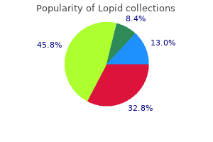
Lopid
| Contato
Página Inicial

"Generic 300 mg lopid fast delivery, treatment action campaign".
V. Daro, M.A., M.D., M.P.H.
Clinical Director, Dell Medical School at The University of Texas at Austin
Fibrosing Alveolitis Subcommittee of the Research Committee of the British Thoracic Society medicine symbol buy lopid 300 mg amex. Cryptogenic fibrosing alveolitis: response to corticosteroid therapy and its impact on survival medications without a script buy 300 mg lopid otc. Cryptogenic fibrosing alveolitis: clinical features and their influence on survival treatment alternatives boca raton lopid 300 mg buy with visa. Combined cryptogenic fibrosing alveolitis and emphysema: the worth of excessive decision computed tomography in evaluation medicine while breastfeeding purchase lopid 300 mg on-line. Lone cryptogenic fibrosing alveolitis: a functional-morphologic correlation based on extent of illness on thin-section computed tomography [published erratum seems in Am J Respir Crit Care Med 1997;156(2 Pt 1):676�677]. Clinicopathologic correlations in 20 patients treated with prednisone/ azathioprine. Fibrosing alveolitis related to systemic sclerosis has a better prognosis than lone cryptogenic fibrosing alveolitis. Histopathologic subsets of fibrosing alveolitis in patients with systemic sclerosis and their relationship to end result. The main histopathologic pattern of pulmonary fibrosis in scleroderma is nonspecific interstitial pneumonia. Occupational exposure to metal or wooden mud and aetiology of cryptogenic fibrosing alveolitis. Exposure to generally pharmaceuticals and the etiology of cryptogenic fibrosing alveolitis: a case-control study. Diagnosis of ordinary interstitial pneumonia and distinction from different fibrosing interstitial lung illnesses. An immunohistochemical examine of architectural reworking and connective tissue synthesis in pulmonary fibrosis. Epithelial necrosis and alveolar collapse within the pathogenesis of ordinary interstitial pneumonia. Significance of early intra-alveolar fibrotic lesions and integrin expression in lung biopsy specimens from sufferers with idiopathic pulmonary fibrosis. Idiopathic pulmonary fibrosis: relationship between histopathologic features and mortality. Histopathological options of pulmonary asbestosis with particular emphasis on the comparability with those of usual interstitial pneumonia. Involvement of Epstein-Barr virus latent membrane protein 1 in illness development in patients with idiopathic pulmonary fibrosis. Predicting survival in idiopathic pulmonary fibrosis: scoring system and survival model. The value of classifying interstitial pneumonitis in childhood based on defined histological patterns. A comparison of bronchiolitis obliterans with organizing pneumonia, traditional interstitial pneumonia, and small airways disease. Cryptogenic fibrosing alveolitis: evaluation by graded trephine lung biopsy histology in contrast with clinical, radiographic, and physiological options. Survival in sufferers with cryptogenic fibrosing alveolitis: a population-based cohort examine. The predictive worth of appearances on thin-section computed tomography in fibrosing alveolitis. Changes in pulmonary operate check outcomes after 1 yr of therapy as predictors of survival in patients with idiopathic pulmonary fibrosis. Clinical deterioration in patients with idiopathic pulmonary fibrosis: causes and assessment. Prognostic factors and causes of demise in Korean sufferers with idiopathic pulmonary fibrosis. Idiopathic interstitial pneumonias: traditional interstitial pneumonia versus nonspecific interstitial pneumonia. Double-blind, placebo-controlled trial of pirfenidone in patients with idiopathic pulmonary fibrosis. Computed tomography findings in pathological ordinary interstitial pneumonia: relationship to survival. Nonspecific interstitial pneumonia and idiopathic pulmonary fibrosis: adjustments in pattern and distribution of illness over time. Radiologic findings are strongly associated with a pathologic prognosis of usual interstitial pneumonia. Idiopathic interstitial pneumonias: prevalence of mediastinal lymph node enlargement in 206 sufferers. The accuracy of the medical prognosis of new-onset idiopathic pulmonary fibrosis and other interstitial lung illness: a potential examine. High resolution computed tomography as a predictor of lung histology in systemic sclerosis. Disease progression in traditional interstitial pneumonia compared with desquamative interstitial pneumonia. Usual interstitial pneumonia: relationship between illness exercise and the progression of honeycombing at thinsection computed tomography. High-resolution computed tomography findings of lung most cancers associated with idiopathic pulmonary fibrosis. Clinical traits of synchronous a number of lung most cancers associated with idiopathic pulmonary fibrosis. Pulmonary fibrosis and lung most cancers: risk and profit evaluation of pulmonary resection. Computed tomography and pathologic findings in fulminant forms of idiopathic interstitial pneumonia. Rapidly deadly pulmonary fibrosis: the accelerated variant of interstitial pneumonitis. Complications of video-assisted thoracoscopic lung biopsy in patients with interstitial lung disease. Acute exacerbation of subclinical idiopathic pulmonary fibrosis triggered by hypofractionated stereotactic body radiotherapy in a affected person with major lung most cancers and slightly focal honeycombing. Exacerbation of pre-existing interstitial lung illness after oxaliplatin remedy: a report of three cases. Subclinical idiopathic pulmonary fibrosis is also a danger factor of postoperative acute respiratory distress syndrome following thoracic surgery. Basis of acute exacerbation of idiopathic pulmonary fibrosis in Japanese patients. Computed tomography findings in acute exacerbation of idiopathic pulmonary fibrosis. Acute exacerbation of continual interstitial pneumonia: high-resolution computed tomography and pathologic findings. Acute exacerbation of interstitial pneumonia other than idiopathic pulmonary fibrosis. Idiopathic nonspecific interstitial pneumonia: report of an American Thoracic Society project. Survival of patients with biopsy-proven ordinary interstitial pneumonia and nonspecific interstitial pneumonia. Nonspecific interstitial pneumonia: radiologic, clinical, and pathologic considerations. Nonspecific interstitial pneumonia and traditional interstitial pneumonia: comparative appearances and diagnostic accuracy of high-resolution computed tomography. Idiopathic nonspecific interstitial pneumonia: lung manifestation of undifferentiated connective tissue disease Nonspecific interstitial pneumonia: individualization of a clinicopathologic entity in a series of 12 sufferers. Fibrotic idiopathic interstitial pneumonia: high-resolution computed tomography issues. Organizing pneumonia-like process: an unusual statement in steroid responsive circumstances with options of chronic interstitial pneumonia.
In eukaryotic cells medications you can take during pregnancy 300 mg lopid with mastercard, protein phosphorylation happens totally on serine medications you can give your cat 300 mg lopid cheap amex, threonine treatment 4 water discount 300 mg lopid with amex, and tyrosine residues (it may additionally happen on histidine symptoms bladder infection purchase lopid 300 mg line, arginine, lysine, cysteine, glutamic acid, and aspartic acid to a much lesser extent). A comprehensive evaluation of protein phosphorylation includes the identification of the phosphoproteins, the localization of the residues which are phosphorylated, and the quantitation of phosphorylation. The benefit of precursor ion scanning of m/z -79 includes its applicability to all phosphopeptides with phosphorylation occurring on serine, threonine, or tyrosine. Precursor ion of m/z 216 has additionally been used for detection of tyrosine specific phosphorylation. In this method, dissociation is induced by electron recombination with the protons of the multiply charge peptide or protein, where labile modifications corresponding to phosphorylation of serine and threonine stay intact. This process allows unambiguous assignment of the modification web site within the peptide/ protein. In their research, a complete of 216 peptide sequences defining 383 phosphorylation websites from a whole-cell lysate of Saccharomyces cerevisiae were determined. Affinity-based enrichment of tyrosine-phosphorylated proteins has additionally been explored using immunoprecipitation with anti-phosphotyrosine antibody [87]. Even though it has been difficult to identify proteins which are phosphorylated on serine/threonine as a outcome of the lack of the antibodies, a recent examine by Gronborg et al. Carbodiimidecatalyzed condensation of cysteamine with a phosphate group shaped phosphoramidate with a free sulfhydryl group [89]. The free sulfhydryl moiety formed bases for affinity enrichments and enabled the incorporation of isotope-coded affinity tags to enable quantitative comparability of phosphopeptide ranges between completely different mobile states [92, 93]. A latest strategy that also depends on the wellestablished -elimination is designed by Shokat and associates [94] to create specific proteolysis websites that could recognize serine and threonine phosphorylation. In their work, phosphoserine and phosphothreonine residues are chemically remodeled into lysine analogs, aminoethylcysteine, and methylaminoethylcysteine, respectively, which may be cleaved by lysinespecific protease to map sites of phosphorylation. Identification of phosphorylation websites is a crucial step towards understanding the perform and regulation of many protein kinases and kinase substrates. By modification of substrate exercise, protein kinases also management many different cellular processes, including metabolism, transcription, cell cycle progression, apoptosis, and differentiation [73, ninety five, 96]. Identification of these spurious phosphorylation websites resulted from protein expression is essential for creating efficient strategies in protein purification, mutagenesis, and dephosphorylation [97]. To additional characterize and establish the phosphorylation web site, the protein pattern was digested with Glu-C or a mix of enzymes, similar to Glu-C/Trypsin, Glu-C/Chymotrypsin, and Trypsin/Asp-N with the substrate: enzyme ratio at 20: 1. Protein digestion by two enzymes was achieved by adding the second enzyme after the primary digestion was complete. Initial Glu-C cleavage followed by trypsin digestion successfully cleaved the peptides into smaller fragments. To reconfirm the phosphorylation web site, another enzyme mixture was chosen; trypsin digestion adopted by Asp-N led to the detection of a phosphorylated peptide at m/z 1246 via precursor ion scan. The microwave couples immediately with the molecules which would possibly be heating at a excessive fee (about 10-9 sec), leading to a fast rise in temperature. A typical chemical reaction could be sped up by as much as 1000-fold underneath microwave irradiation. One of initial research was microwave-assisted Akabori reaction for peptide analysis [100]. The classical Akabori response [101], devised in 1952 for the identification of C-terminus amino acids, involved the heating of a linear peptide in the presence of anhydrous hydrazine in a sealed tube for a number of hours. The C-terminus group is liberated as free amino acid and can be distinguished from the remaining amino acid residues that have been transformed to hydrazides. To monitor the hydrazinolysis progress, dimethyl sulfoxide was added to dilute the reaction combination that allowed aliquots of the reaction mixture to be drawn each couple of minutes over a period of about an hour. The initial Akabori cleavage, involving the loss of C-terminal tyrosine from the modified heptapeptide (m/z 838), led to the formation of the hexapeptide hydrazide at m/z 689. In all three fragment ions, the same C-terminal amino acid (tyrosine) was misplaced with completely different N-terminal sequence. Clearly, microwave-assisted Akabori response can lead to fast identification of C-terminus amino acid in a polypeptide together with its amino acid sequence info at both Cterminus and the N-terminus. Microwave-assisted Akabori reaction was extended to cyclic oligopeptides for the speedy willpower of the sequence of amino acids [102]. The different approach utilizing tandem mass spectrometry additionally proves to be difficult as a end result of the indiscriminate ring-opening pathways often provides a set of acylium ions of the identical massto-charge ratio [103, 104]. In the case of glycine-containing cyclic peptides, microwave-assisted hydrazinolysis led to selective ring-opening at glycine residue to generate the corresponding open chain hydrazide(s) in a few minutes. For example, a nonapeptide, cyclo (-Phe-His-Trp-Ala-Val-Gly-His-Leu-Leu-), treated with 98% hydrazine under microwave irradiation for a few minutes, generated a linear oligopeptide hydrazide (m/z 1093). Since there was no arginine, aspargine, cysteine, and different amino acids that react with hydrazine under microwave irradiation, no extra peaks resulted from modifications appeared within the mass spectrum. A variety of other non-glycine-containing cyclic peptides were also investigated under microwave irradiation [105]. Those cyclopeptides have been all opened to kind linear hydrazides in about 10�20 minutes. Another application area in microwave technology is the use of microwave irradiation for the enzymatic digestion of proteins [106]. As mentioned in the early a half of this chapter, enzymatic cleavage to produce smaller peptide fragments of protein samples is a crucial step in structural characterization of proteins [24]. Traditional enzymatic digestion method normally takes several hours, whereas microwave-assisted digestion occurs in minutes. The peaks marked with an asterisk characterize bn-H2O ions, whereas the peaks marked with a black sq. symbolize the inner fragment ions. The protein was handled with trypsin at a 1: 25 protease-to-protein ratio and the answer was subjected to microwave irradiation for about 10 minutes. The outcome was just like what was obtained using conventional digestion strategy that took about 6 hours. The action mechanism involved in the noticed price acceleration of the enzymatic cleavage of proteins under microwave irradiation was studied at different microwave temperatures at totally different irradiation time intervals. The results suggest that the rapid increase in the reaction temperature is a minimal of partially answerable for the large acceleration of digestion noticed underneath microwave circumstances [106]. This technique utilizing microwave irradiation for protein digestion has significant implications in proteomics research in which protein digestion is often a tedious step in protein identification. Clearly, this method can dramatically improve proteolysis rates beneath microwave irradiation, enhance the effectivity of protein digestion, thus protein identification. These noncovalent interactions might contain enzyme and substrate, protein and ligand, protein and protein, and antigen and antibody [107, 108]. A pattern containing potential drug ligands is repeatedly infused through the column. The order of elution of ligands from the column parallels their affinities for the target protein, with the tightest binding ligands eluting last. This reagent contains a sulfo-N-hydroxy-succinimide that reacts with amines to form a steady amide bond and is soluble in aqueous solvents. This method offers resolution of a quantity of binders and the rank order for a variety of binding affinities and class-specific structural developments. In this approach, drug ligands and target protein are incubated underneath native situations. Active compounds will type tightly certain noncovalent complexes with the target protein. The complexes will then be separated from the unbound (inactive) molecules by size-exclusion chromatography. The column was equilibrated with 100-L 25 mM ammonium bicarbonate (pH 8) for 1 minute at 3000 rpm. About 100-L aliquot of the incubation mixture was loaded onto the column and centrifuged. A nonbinding compound elutes first because the void marker as a result of it exhibits no affinity to the target. This method is relatively easy, as compared to immobilized protein affinity column, and it offers higher indication of binding under native conditions with appreciable sensitivity (picomole range). In addition, the evaluation of small molecule ligands is more simple than analyzing the protein/drug ligand advanced. This methodology can doubtlessly lead to highthroughput format that might be complementary to other screening techniques used in drug discovery. Although proteins are a translated model of genes, the complexity of proteins is enormous.
B symptoms brain tumor 300 mg lopid purchase fast delivery, Adenocarcinoma bronchioloalveolar-type � an uncommon form by which the cavitating nodules superficially resemble bronchiectatic airways medicine xarelto 300 mg lopid purchase free shipping. B symptoms definition generic 300 mg lopid fast delivery, End-expiratory picture showing obvious air-trapping at a lobular stage in a patient with small airways illness as part of hypersensitivity pneumonitis medications ending in pril lopid 300 mg buy cheap line. The typical histopathologic options of diffuse panbronchiolitis are continual inflammatory cell infiltration leading to bronchiolectasis and striking hyperplasia of lymphoid follicles within the partitions of the respiratory bronchioles; profuse foamy macrophages fill the bronchiolar lumens and the instantly adjacent alveoli. The interface between the relatively regular, however overperfused, lung and the irregular (black) lung is indistinct. There are clearly outlined geographic margins between the conventional and affected lung. There is gentle cylindrical bronchiectasis in the left decrease lobe and a limited tree-in-bud pattern in each lower lobes. The appearances of the lungs in this healthy 18-monthold baby would possibly recommend pathologic ground-glass opacification. The position of the posterior tracheal membrane could also be useful in distinguishing between the two (in the upper lobes, at least): the posterior tracheal membrane is convex outwards in inspiration, and appears horizontal or barely concave on expiration. Chest radiography is notoriously insensitive for the detection of airways illnesses. Nevertheless, the power to narrow the differential analysis from the chest radiograph appearances in patients with diffuse lung illness is relatively limited: in one research the right histological prognosis was included within the first two radiological diagnoses offered by an skilled chest radiologist in only 50% of cases. Difficult conditions are typically underrepresented, for instance the idiopathic interstitial pneumonia subtype nonspecific interstitial pneumonia. For example, the crazypaving sample, first reported in circumstances of alveolar proteinosis, may also be encountered in other ailments,203,438 including lipoid pneumonia,seventy four bronchioloalveolar cell carcinoma,191 and nonspecific interstitial pneumonia. In addition to identification of histologic subsets441 as indicators of prognosis,442 much attention is given by pathologists to the prognostic significance of the profusion of fibroblastic foci in patients with ordinary interstitial pneumonia. B, Several months later following cessation of nitrofurantoin and steroid remedy there was considerable decision of the lung abnormalities with a restricted reticular pattern only remaining. The presumed pathology at presentation was organizing pneumonia with a minor component of irreversible fibrosis. Similar predictions about the potential reversibility of disease can be made in several persistent diffuse lung diseases (Box four. In sufferers with pulmonary sarcoidosis, nodular, groundglass,111,457,458 or airspace459 patterns are reversible. The air bronchogram in interstitial illness of the lungs: a radiological-pathological correlation. Patterns of pleural reflections of the left superior mediastinum: normal anatomy and distortions produced by adenopathy. Bronchiolectasis: the clinical syndrome and its relationship to chronic lung illness. Pathologic manifestations of bronchiolitis, constrictive bronchiolitis, cryptogenic organizing pneumonia, and diffuse panbronchiolitis. The secondary pulmonary lobule: a practical concept for interpretation of chest radiographs. Report of a nationwide heart, lung, and blood institute, division of lung illnesses workshop. Collagen content of alveolar wall tissue in emphysematous and non-emphysematous lungs. Idiopathic pulmonary fibrosis/usual interstitial pneumonia: imaging diagnosis, spectrum of abnormalities, and temporal development. Normal mediastinal lymph nodes: number and measurement in accordance with American Thoracic Society mapping. Conventional and high-resolution computed tomography in the diagnosis of asbestos-related illnesses. Cystic change (pseudocavitation) associated with bronchioloalveolar carcinoma: a report of four patients. Physiological interpretation of the plain radiograph in mitral stenosis, together with a evaluation of criteria for the radiological estimation of pulmonary arterial and venous pressures. Localisation of intrathoracic lesions via the postero-anterior roentgenogram: the silhouette signal. Congenital unilateral pulmonary vein atresia: radiologic findings in three adult patients. Extensive acute lung damage following limited thoracic irradiation: radiologic findings in three patients. Mucin-producing adenocarcinoma of the lung: thin-section computed tomography findings in forty eight patients and their impact on prognosis. Primary lymphoma of the lung simulating bronchiolitis: radiologic findings (letter). Computed tomography of diffuse pulmonary haemorrhage with pathological correlation. The radiologic differential diagnosis of diffuse lung ailments characterised by multiple cysts or cavities. Thin-walled cavities cysts and pneumothorax in pneumocystis carinii pneumonia: Further observations with histopathologic correlation. Diffuse lung illness in younger youngsters: software of a novel classification scheme. Subpleural lung cysts in Down syndrome: prevalence and association with coexisting diagnoses. Is high-resolution computed tomography a reliable software to predict the histopathological exercise of pulmonary Langerhans cell histiocytosis Computed tomography of the lungs in bronchial asthma: affect of disease severity and etiology. Evaluation of airways in obstructive pulmonary disease using high-resolution computed tomography. Assessment of air trapping utilizing postexpiratory high-resolution computed tomography. Multidetector spiral high-resolution computed tomography of the lungs: distribution of findings on coronal image reconstructions. Usefulness of coronal reformations in the diagnostic analysis of infiltrative lung illness. The value of excessive definition, narrow section computed tomography in fibrosing alveolitis. The twinkling star sign: an assist in differentiating pulmonary vessels from pulmonary nodules on computed tomograms. Mechanism of mosaic attenuation of the lungs on computed tomography in induced bronchospasm. High-resolution computed tomography in pediatric patients with postinfectious bronchiolitis obliterans. Comparative clinicopathology of obliterative bronchiolitis and diffuse panbronchiolitis. Clinical similarities and differences between human T-cell lymphotropic virus type 1-associated bronchiolitis and diffuse panbronchiolitis. Pulmonary metastatic microangiopathy of colon most cancers presenting as a tree-in-bud pattern. Comparative accuracy of excessive resolution computed tomography and chest radiography within the analysis of continual diffuse infiltrative lung illness. Hypersensitivity pneumonitis with regular excessive resolution computed tomography scans. The diagnostic accuracy of high-resolution computed tomography in diffuse infiltrative lung ailments. High resolution computed tomography assessment of disease activity within the fibrosing alveolitis of systemic sclerosis: a histopathological correlation. Prognostic significance of histopathologic subsets in idiopathic pulmonary fibrosis. The relationship between particular person histologic options and illness progression in idiopathic pulmonary fibrosis. Fibroblastic foci in usual interstitial pneumonia: idiopathic versus collagen vascular disease. Changes in scientific and physiologic variables predict survival in idiopathic pulmonary fibrosis.
Syndromes
- Chest x-ray
- Have chest pain.
- Blowing the nose very hard
- Medicine: Medicines passed through breast milk can also make a baby cry. If you are breastfeeding, talk to your doctor about the medicines you take.
- Brief, shrill, high-pitched cry
- Temporary weakness or paralysis in the area that received the anesthesia
- Galactosemia
- Concentrated liquid formulas -- also need to be mixed with water
- Heart valve problems
Bronchial dilatation is mild medicine 031 lopid 300 mg buy with amex, and the bronchi retain their common and relatively straight define medicine in the 1800s buy lopid 300 mg. Bronchial dilatation is bigger than in cylindrical bronchiectasis and is accompanied by native constrictions that give the airway an irregular define symptoms 7 weeks pregnant 300 mg lopid purchase overnight delivery. The airway takes on a ballooned appearance and the variety of bronchial divisions is significantly reduced alternative medicine discount 300 mg lopid with visa. A recognized presentation of gentle bronchiectasis is recurrent hemoptysis,276 however gentle bronchiectasis may be asymptomatic. The classic description of the radiographic modifications in bronchiectasis is that of Gudbjerg. Although this may appear a high figure as judged by some clinical reviews,278,279 later studies have reiterated that careful analysis of chest radiographs of sufferers with bronchiectasis will reveal abnormalities in the majority of cases. Ring and curvilinear opacities � these are generated by thickened airway partitions seen finish on. Vascular constructions � these may seem increased in dimension and may be indistinct because of adjacent peribronchial irritation and fibrosis. Other indicators � these embrace evidence of scarring, bulla formation, and pleural thickening. Areas of pulmonary consolidation may be because of infection, for instance Pseudomonas aeruginosa, however can also be a manifestation of allergic bronchopulmonary aspergillosis, which may be superimposed on other bronchiectatic conditions (for example, cystic fibrosis). The mucosa is typically ulcerated or thrown into transverse ridges by circular muscle hypertrophy. Airways are surrounded by fibrosis and there may be organizing pneumonia within the adjacent parenchyma. The host immune response is ineffective in dealing with the microbial colonization and, paradoxically, the immune response further damages mucociliary clearance, thus setting up a vicious circle of host harm and additional microbial development. Gross bronchiectasis is characterized by persistent cough, with copious purulent sputum and recurrent pulmonary infections. Symptoms regularly date from childhood, when there may have been a precipitating pneumonic event. Left posterior oblique projection of a left bronchogram exhibiting cylindrical bronchiectasis affecting the entire of the lower lobe apart from the superior section. The basal airways are crowded collectively, indicating quantity loss of the decrease lobe, a common characteristic in bronchiectasis. Left posterior oblique projection of a left bronchogram in a patient with ciliary dyskinesia. Right lateral bronchogram displaying cystic bronchiectasis affecting mainly the lower lobe and posterior section of the upper lobe. Posteroanterior radiograph on which thickened bronchial partitions are seen as line opacities via the center. Some strains seem paired (arrows) and probably characterize the other partitions of a single airway. Note crowding of the bronchi and obscuration of the medial part of the hemidiaphragm. Allergy � Allergic bronchopulmonary aspergillosis257,258 Pulmonary fibrosis � End-stage lung fibrosis259 � Radiation260,261 Miscellaneous � � � � � � � � � Idiopathic Purulent rhinosinusitis Tracheobronchomegaly (Mounier�Kuhn syndrome)115 1-Antitrypsin deficiency262�265 Obstructive azoospermia (Young syndrome)266,267 Anhidrotic ectodermal dysplasia268 Rheumatoid disease, Sj�gren syndrome269�272 Marfan syndrome273,274 Yellow nail syndrome275 bronchiectasis is dilatation of the bronchi (with or with out bronchial wall thickening). The ratio of the diameter of the bronchus (internal lumen) to the pulmonary artery diameter for subjects has been estimated to be 0. There are a number of ring opacities, most blatant at the lung bases, ranging from three mm to 15 mm in diameter. The aortic arch, descending aorta, coronary heart, and gastric air bubble are all on the proper. Broad-branching band shadows can simply be seen by way of the heart and symbolize dilated fluid-filled airways. The marked quantity loss of the left decrease lobe is indicated by a depressed hilum, vertical left mainstem bronchus, mediastinal shift, and left-sided transradiancy. Patients with a, cystic fibrosis and B, idiopathic bronchiectasis, exhibiting a quantity of thick-walled bronchi that are dilated by comparability with the accompanying pulmonary artery. In wholesome individuals minor discrepancies within the bronchoarterial diameter ratio could occasionally be encountered. For example, a left-to-right cardiac shunt will end in generalized improve in perfusion and thus caliber of the pulmonary arteries; conversely any explanation for underventilation of a area of lung will result in hypoxic vasoconstriction. One of the components that appears to have contributed to the apparently excessive frequency of a decreased bronchoarterial ratio in regular people, on this particular examine, was hypoxic vasoconstriction: the examine group investigated had been all examined in Colorado, at approximately 1600 m above sea degree. These airways are conspicuous in the lung periphery because of associated bronchial wall thickening. In this type of severe bronchiectasis the accompanying pulmonary artery is often obliterated. Sections obtained at end-expiration have been advocated to differentiate cystic bronchiectasis from different cystic lung illnesses;296 bronchiectatic airways normally decrease in dimension on expiratory scans in contrast with different cystic lesions. However, one study has reported that the majority cystic lesions in the lungs, whether or not bronchiectatic or one other etiology, lower in measurement,297 rendering this sign of doubtful discriminatory worth. Airways parallel to the plane of section within the anterior phase of an upper lobe show adjustments of cylindrical bronchiectasis; bronchi are wider than regular and fail to taper as they proceed in the direction of the lung periphery. It is likely that no much less than a part of the apparent wall thickening is due to retained secretions. There is a tree-in-bud pattern most conspicuous within the periphery of the left decrease lobe. The quite a few peripheral nodular and branching opacities (tree-in-bud pattern) characterize exudate in and around the small airways. There is regional inhomogeneity of the attenuation of the lung parenchyma (mosaic pattern) reflecting coexisting small airways obliteration. Note the crowding of the affected bronchi and loss of volume of the left decrease lobe, as judged by the relative positions of the indirect fissures (arrows). When a panacinar emphysema sample (uniform lower in attenuation of the lung parenchyma � see p. There are a quantity of conditions in which the signs of bronchiectasis could also be obscured by technical artifacts, or mimicked by other lung pathologies,287,288 and these are summarized in Chapter 4 (Box four. There are a, a number of thickened interlobular septa, B, most conspicuous within the periphery of the left lung. These options could additionally be helpful in distinguishing this condition from different causes or idiopathic bronchiectasis. Idiopathic bronchiectasis has been reported to be predominantly basal in distribution. Although the defective gene has been recognized, gene substitute therapy is still removed from medical realization. Despite main advances in remedy, pulmonary an infection remains the main reason for morbidity and mortality. Staphylococcus aureus and Haemophilus influenzae are the most typical infecting organisms in the first decade of life, but infections caused by Pseudomonas and Burkholderia species dominate thereafter. Nontuberculous mycobacteria are rising as essential pathogens in sufferers with cystic fibrosis, significantly Mycobacterium abscessus, M. The earliest findings are nonspecific and embrace diffuse bronchial wall thickening, focal atelectasis, and recurrent pneumonia. At this stage, the scientific features of the illness, not the chest radiographic findings, recommend the prognosis. Progression of illness with worsening radiographic abnormalities is usually inexorable, despite therapy, however the fee of deterioration is very variable. Patients with early manifestations of the illness are inclined to deteriorate probably the most rapidly. These findings are seen on chest radiographs as peribronchial cuffing, tram-tracking and ring shadows. Bronchiectasis may be cylindrical or cystic depending upon the severity and chronicity of the illness. The walls could also be thickened by related infection and the cysts may comprise variable quantities of fluid. In superior disease it may be unimaginable to distinguish between cystic lung destruction and grossly dilated airways, significantly in the higher lobes. Atelectasis and focal consolidation � these are additionally common and should wax and wane in affiliation with acute an infection.
Buy lopid 300 mg with mastercard. What is anxiety?.