
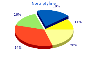
Nortriptyline
| Contato
Página Inicial

"Nortriptyline 25 mg discount on line, anxiety while sleeping".
R. Rakus, M.B. B.CH. B.A.O., M.B.B.Ch., Ph.D.
Professor, Eastern Virginia Medical School
Normal persons who devour a typical Western diet ingest approximately 70 mmol of potassium per day anxiety getting worse nortriptyline 25 mg cheap with amex. The small intestines anxiety symptoms hives nortriptyline 25 mg buy without prescription, particularly jejunum anxiety 1 mg cheap nortriptyline 25 mg without prescription, absorb virtually all the ingested potassium and ship it to the liver for processing by means of the hepatoportal circulation anxiety symptoms throat closing order 25 mg nortriptyline fast delivery. In a balanced state of potassium, eighty to eighty five p.c of day by day consumption of potassium is excreted by the kidneys. The remaining quantity of potassium consumption is mostly excreted by the gastrointestinal tract. Potassium is filtered by the glomeruli and virtually utterly reabsorbed by the proximal tubule and ascending limbs of loop of Henle. Potassium is secreted by the principal cells of the accumulating tubules, and most of this potassium is excreted within the urine. Factors that regulate principal cell potassium secretion embody potassium consumption, intracellular potassium level, sodium delivery to the amassing tubules, urine move price, and hormones, such as aldosterone and betacatecholamines. The kidneys are the main organs for exterior potassium balance or homeostasis; therefore, extreme Hypokalemia: Prevention and Treatment forty seven lack of potassium via the kidneys eventually leads of hypokalemia. This mechanism minimizes the postprandial increase in plasma potassium focus. To keep stability, the amount of potassium consumed within the meal (minus the small amount misplaced in the feces) is secreted into the urine. This pathway of potassium loss can be a crucial supply of safety towards hyperkalemia when renal operate is severely decreased. Conversely, if potassium intake is low or its output could be very high, plasma potassium concentration decreases and suggestions regulation redistributes potassium from intracellular fluid to plasma and minimizes renal potassium excretion. Insulin Insulin stimulates intracellular uptake of potassium and might quickly scale back extracellular potassium focus. This is likely to occur after a excessive carbohydrate meal in individuals with normal pancreas. Conversely, in diabetes mellitus the deficiency of insulin will enhance the propensity to hyperkalemia. However, this proneness to hyperkalemia is mitigated by the osmotic diuresis and accompanying kaliuresis in uncontrolled diabetes mellitus. Catecholamines Among the catecholamines, epinephrine influences inner potassium influx and produces hypokalemia without growing urinary excretion of potassium, suggesting shift of potassium from the extracellular fluid to intracellular fluid. Hypokalemic impact of epinephrine could be demonstrated by infusion of epinephrine into man or animal. The significance of intracellular translocation of potassium induced by epinephrine in regular circumstances is unclear; however, this impact presents a doubtlessly protective mechanism in opposition to life-threatening hyperkalemia in patients with large trauma, postsurgery, or rhabdomyolysis. Each of these conditions could result in an enormous release of intracellular potassium. With decreased function of the kidneys, ability to excrete potassium load is diminished, however internal shift of potassium provides a safety against dangerous hyperkalemia in states of potassium overload. After consideration of the elements that management exterior and inner potassium homeostasis, the causes of hypokalemia could be appropriately defined. It is necessary to reiterate that amongst all these components, high renal excretion of potassium determines most cases of hypokalemia. In metabolic alkalosis, although hypokalemia has been claimed to be as a outcome of alterations in internal potassium balance, urinary loss of potassium may account for a large a half of hypokalemia. Both acute and persistent respiratory alkalosis has slight effect on inner or exterior potassium balance with a gentle tendency to hypokalemia. The Common Causes of Hypokalemia Diuretic Therapy Diuretic remedy is the commonest explanation for hypokalemia of all severity. All diuretics-whether thiazides, loop diuretics, or carbonic anhydrase inhibitors-produce hypokalemia of variable severity. Thus, immediate look of hypokalemia following diuretic remedy is an indication of a preexisting covert renal potassium leak condition, such as primary or secondary hyperaldosteronism. Alcoholism Alcoholics delivered to the emergency room reveal a panorama of metabolic issues, consisting of hypokalemia, hypophosphatemia, and hypomagnesemia. Hypokalemia is the most common abnormality found and is due to a number of elements, together with poor intake of potassium, gastric loss of potassium by way of vomiting, and urinary lack of potassium. Severe Vomiting and Gastrointestinal Suction and Drainage Patients with severe vomiting or gastrointestinal suction and drainage following surgical procedure regularly develop severe hypokalemia. Hypokalemia is partly due to gastric loss of potassium and partly due to renal loss of potassium. A sequence of occasions begins with volume depletion and resulting in hyperreninemia and hyperaldosteronism that accounts for the renal loss of potassium. Severe Diarrhea Severe diarrhea is changing into a typical cause of extreme hypokalemia within the hospital setting. The extent of K loss in the diarrheal stool is difficult to decide as no methodology is available. Primary or Secondary Hyperaldosteronism Hypokalemia in a hypertensive patient in the absence of diuretic use should alert a doctor about the chance of major aldosteronism (aldosterone producing tumor, bilateral adrenal hyperplasia) or secondary hyperaldosteronism due to renal artery stenosis or reninsecreting tumor. The Uncommon Causes of Hypokalemia Leukemia Moderate to extreme hypokalemia is very common in acute myelocytic or myelomonocytic leukemia. The actual mechanism(s) by which lysozyme excretion induces kaliuresis and hypokalemia are unknown. Hypokalemia Associated with Ectopic Adrenocorticotrophic Hormone Production Tumors, particularly small cell-type bronchogenic carcinoma, might produce an adrenocorticotrophic hormone that stimulates cortisol and aldosterone production and, thereby, enhances urinary potassium loss. Hypokalemia could be intractable and would require giant amounts of potassium supplement to management hypokalemia. Hypokalemia Induced by Antibiotic Many antibiotics, together with penicillin, carbenicillin, and gentamicin, produce hypokalemia. Hypokalemia is as a result of of extreme secretion of potassium, which happens in order to dissipate luminal electronegativity caused by penicillin or carbenicillin. Renal cortical tissue after gentamicin administration demonstrates extreme numbers of lysosome. These lsosomes have been found to be the storage supply of lysozyme, resulting in lysozymuria and consequent hypokalemia. Magnesium Depletion Hypomagnesemia is a concomitant abnormality with hypokalemia and is seen commonly in sufferers treated with a loop diuretic, with malabsorption syndrome, and in alcoholics. The mechanism(s) by which hypomagnesemia causes hypokalemia is unknown; however, it ought to be confused that hypomagnesemia might delay correction of hypokalemia with only potassium supplementation. Short stature, failure to thrive, muscle weak spot or cramps, polyuria and nocturia are the usual presenting options. The attribute features embrace hypokalemia, hyperreninemia, and hyperaldosteronism. The supply of enormous quantities of sodium and chloride in the distal and collecting tubules could, partly, be liable for the excessive potassium secretion and kaliuresis. Defect in sodium and chloride reabsorption outcomes also in lack of a cotransporter, calcium. This syndrome contains low plasma renin activity, low plasma aldosterone level, and elevation of blood strain. This syndrome apparently is the outcome of an unknown circulating mineralocorticoid and, consequently, hypervolemia from Na+ and water retention. The situation is characterised by intermittent sudden episodes of severe muscle weakness to paralysis of the lower extremities or of all four extremities. Hypokalemia is due to an abnormality within the inside potassium homeostasis resulting in rapid shift of potassium from the extracellular fluid into the intracellular fluid. These episodes are precipitated by a high carbohydrate meal, glucose, insulin, epinephrine, and even severe anxiety with accompanying respiratory alkalosis, and possible outpouring of epinephrine. Chronic Laxative Use Laxative (licorice) incorporates glycyrrhizic acid, which tends to promote sodium reabsorption resulting in hypervolemia and hypertension because of mineralocorticoid excess. In the state of mineralocorticoid extra, the enzyme that converts cortisol to cortisone is inactivated allowing higher ranges of cortisol. Leukemia Low Intake of Potassium As current medical practice entails increasing number of aged and disabled population in the hospital setting, incidence of hypokalemia is high among the many hospitalized sufferers. Most probably hypokalemia in these population is due to poor consumption of meals that are rich in potassium, such as meat, fruits, fruit juice, and green greens. Poor consumption of potassium coupled with compulsory renal loss of potassium, which is 10 to 15 mmol per day, makes hypokalemia worse. Potassium, being the major intracellular cation, muscular tissues cells are most severely affected in potassium depletion syndrome. Therefore, the manifestations of hypokalemia are largely ascribed to the practical derangements of skeletal and heart muscle tissue and smooth muscles of the gastrointestinal tract.
Syndromes
- Long-term, daily sun exposure (such as in people who work outside)
- Dry cough
- Permanent brain damage
- Breast lump
- Delayed growth and development
- Malignant hypertension (arteriolar nephrosclerosis)
- Hospitalization
- Sick sinus syndrome
- Six servings/day
The mixing of oxygenated and deoxygenated blood that happens on account of an intrapulmonary shunt is called venous admixture anxiety hives discount 25 mg nortriptyline with visa. The time period venous admixture is also used interchangeably in some texts with the time period intrapulmonary shunt ms symptoms anxiety zone order 25 mg nortriptyline amex. Different types of intrapulmonary shunts can happen anxiety icd 9 nortriptyline 25 mg order without a prescription, with the venous admixture being the outcome of these shunts anxiety symptoms perimenopause generic 25 mg nortriptyline mastercard. Absolute shunts, also identified as complete shunts, are made up of the sum of capillary shunts and anatomic shunts. Capillary shunts describe blood that passes by way of the pulmonary capillaries of unventilated alveoli, the systemic blood provide to the airways of the lungs, or deoxygenated blood flowing instantly from pulmonary arterioles to close by pulmonary veins by way of anastomoses, thereby bypassing the alveolar capillaries. A n anatomic shunt is when unoxygenated blood passes via the pulmonary circulation without the chance for gas exchange to occur. An anatomic shunt that impacts 2�3% of the cardiac output happens naturally in healthy individuals. In regular lungs, blood from the bronchial, pleural, and Thebesian veins flows into the pulmonary veins then into the left atrium, thus bypassing the alveoli. This harm or irregularities may be caused by vascular lung tumors or intracardiac shunts. Absolute shunts that occur as a outcome of intracardiac shunts can be categorized as both a right-to-left shunt or a left-to-right shunt. This allows unoxygenated blood to flow from the best side of the center to the left facet of the center. Congenital defects such as transposition of the great vessels, tricuspid atresia, and tetralogy of Fallot are among the many most common causes of right-toleft shunts. This allows blood to flow again into the right heart and to the pulmonary circulation without passing through the systemic circulation. At start, the toddler has a natural rise in PaO2 and a decline in the focus of a lipid called prostaglandin. Under regular conditions, these two components trigger the closure of the ductus arteriosus. In infants with an open ductus, the blood flows from the aorta to the pulmonary artery, leading to a left-to-right shunt. Description Other, less common, kinds of congenital defects that may also cause a leftto-right shunt include ventricular septal defect, which is a hole in the wall of the septum that separates the left and right ventricles; an atrial septal defect, which is a hole within the septum that separates the left and proper atria; or an atrioventricular defect, which is a time period used to describe quite so much of congenital coronary heart malformations. Relative shunting happens when perfusion in the pulmonary capillaries is larger than alveolar air flow. Relative shunting could happen as a end result of alveolar capillary defects, as a response to a sudden improve in the cardiac output, or sudden dilation the pulmonary vasculature. In these circumstances, the blood moves too rapidly previous the alveoli for gasoline exchange to happen, which ends up in insufficient quantities of oxygen diffusion. Shunt Equation the diploma of shunting and venous admixture can be assessed utilizing the shunt equation. It is usually calculated in critically ill pulmonary patients and clinically is recognized as the pulmonary shunt fraction (s/ t). Four parameters have to be determined before the pulmonary shunt fraction calculation could be completed. The first parameter is the arterial partial stress, Pao2, which is calculated using the alveolar gasoline equation. The Pao2 is required to decide the following parameter, the end-pulmonary capillary oxygen content (Cco2). The remaining two parameters are the oxygen content material of arterial blood (CaO2) and the oxygen content material of blended venous blood (C o2). The alveolar fuel equation is as follows: the Pao2 is important to complete the equation for the Cco2. The equation for pulmonary shunt fraction is a mathematical technique for figuring out s based on recognized or calculated variables. A 28-year-old woman with bilateral pulmonary infiltrates on her chest radiograph is admitted to the intensive care unit for shortness of breath. She experiences a respiratory arrest, after which is intubated and placed on mechanical ventilation. Another way to think of the shunt equation is as a comparison of arterial oxygen content material to pulmonary end-capillary oxygen content material. This could additionally be a continual, secure situation, or it might be the early levels of a extra severe situation. A shunt higher than 30% is considered an emergency and requires instant assessment and treatment in most cases. Distribution of Pulmonary Blood Flow Gravity and the place of the physique have direct results on the distribution of blood throughout the pulmonary vascular system. The variations in blood move from the apex of the lungs to the base may be instantly attributed to the stress adjustments that happen on the pulmonary capillary level because of changing place. When an individual stands upright, the distribution of blood from the highest to the bottom of the lungs diminishes proportionally. When the same individual lies down on their again, the amount of blood from the apex of the lungs to the base turns into more evenly distributed. However, because of gravity, the posterior segments of the lungs obtain extra blood than the anterior segments on this position. Dividing the lungs into three zones supplies a better understanding of the effects of gravity on pulmonary blood move. Normally, an individual has enough pulmonary artery stress to present enough blood circulate. However, if the pulmonary artery pressures lower and gravity causes the arterial and venous pulmonary pressures to be decrease than the intra-alveolar pressures when the individual breathes, the larger strain within the alveoli compresses the surrounding capillaries and occludes blood flow, thereby stopping the perfusion. In zone 2, the arterial blood pressures within the pulmonary capillaries are greater than the alveolar pressures; nevertheless, the pulmonary venous pressures are lower than the alveolar pressures. This causes an intermittent blood flow via the pulmonary circulation in zone 2; that is typically known as a waterfall impact. The waterfall impact happens due to the variations between the pulmonary arterial pressures and the intra-alveolar pressures. As the blood flows by way of the pulmonary capillaries from the arterial facet to the venous aspect, the blood stress steadily decreases. At the purpose at which the intra-alveolar pressures become greater than the pressures in the pulmonary capillaries, the capillaries collapse, and blood circulate stops. This creates a brief enhance in pulmonary vascular resistance, and strain begins to build within the capillaries till the capillary strain exceeds the intra-alveolar strain and briefly reopens the capillaries. As the capillaries reopen, the blood gushes as if falling over a waterfall, after which progressively the pressures return to their lower stage, the capillaries collapse, and the process starts all over again. Under perfect circumstances, each space of the lung could be absolutely perfused and fully ventilated. As discussed earlier, a shunt happens when the capillaries collapse and the blood is shifted to betterventilated areas of the lungs. A comparison of the rate of ventilation to the speed of perfusion might help quantify these conditions. It may be calculated by dividing the motion of air measured in liters per minute (ventilation) by the flow of blood measured in liters per minute (perfusion). For example, underneath normal circumstances, the alveolar air flow is 4 L/min and the conventional capillary blood flow is 5 L/min, which yields a ratio of 0. The / ratio instantly affects the absorption of oxygen and the discharge of carbon dioxide between the alveolus and the blood. In the primary mechanism, a volume of fluid strikes from an area of higher hydrostatic stress in the capillary bed to an space of decrease hydrostatic stress within the interstitium. By definition, hydrostatic pressures push fluid from an area of upper focus to an area of decrease concertation; this is known as filtration. Oncotic pressure pulls water molecules from the interstitial house back into the capillary, shifting from an space of low focus to an space of higher concentration; this is known as reabsorption. No internet movement of fluid occurs close to the midpoint because these two pressures are roughly equal at this stage. In this instance from the systemic circulation, the hydrostatic stress in the interstitium (Pi) is 1 mm Hg and the hydrostatic stress in the capillary (Pc) is 45 mm Hg. The fluid is pushed from an area of higher concentration (45 mm Hg) to an area of decrease focus.