
Top Avana
| Contato
Página Inicial
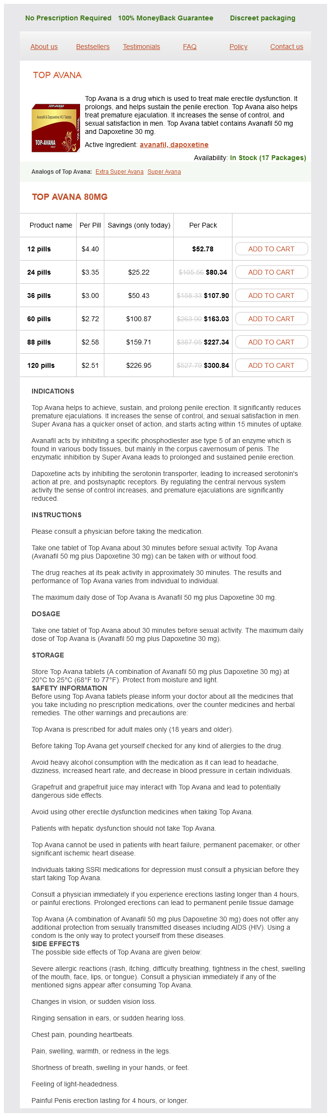
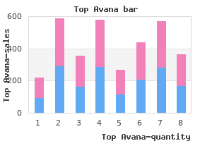
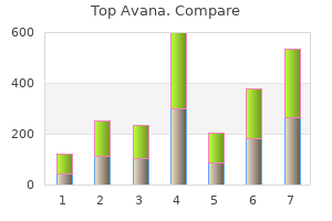
"80 mg top avana buy, erectile dysfunction meds at gnc".
F. Ali, M.B.A., M.D.
Clinical Director, University of Oklahoma College of Medicine
Laryngeal stimulation produces a choking sort of cough without a preceding inspiration erectile dysfunction causes relationship problems purchase top avana 80 mg mastercard. Inadequate mucociliary clearance mechanisms (as in bronchiectasis or cystic fibrosis) may produce a pattern of coughing with less violent acceleration of air and a sequence of interrupted expirations with none intervening inspiration erectile dysfunction pills for high blood pressure purchase top avana 80 mg visa. Many problems can impair the flexibility to cough successfully buy erectile dysfunction injections order 80 mg top avana otc, which can result in persistent cough erectile dysfunction protocol video buy discount top avana 80 mg on-line. The aged, the new child, lung transplant recipients and sufferers with paralysis or neuromuscular issues have a poorly developed and/or compromised cough reflex, and are rendered highly vulnerable to infective and aspiration pneumonia (Polverino et al 2012). Individuals with decreased function of the stomach wall musculature are particularly vulnerable to ineffective cough. Individuals with laryngeal disorders, together with those with tracheostomies, may not achieve adequate laryngeal closure to generate the elevated intrathoracic pressures necessary for an effective cough. They may be differentiated from other airway afferents by their rapid adaptation (in 1�2 seconds) to sustained lung inflation (Schelegle and Green 2001). Their activation initiates reflex bronchospasm and mucus secretion through parasympathetic pathways. Their exercise increases throughout inspiration and peaks just prior to the initiation of expiration. They are thus thought to be the afferent fibres concerned within the Hering�Breuer reflex, which terminates inspiration and initiates expiration when the lungs are adequately inflated. C fibres nearly all of afferent nerves innervating the airways and lungs are unmyelinated C fibres, just like the unmyelinated somatic sensory nerves that innervate the pores and skin, skeletal muscle, joints and bones and that reply to noxious chemical and mechanical stimuli. They are usually quiescent throughout the respiratory cycle (Lee and Pisarri 2001). Ciaglia P, Firsching R, Syniec C 1985 Elective percutaneous dilatational tracheostomy. Nabati M, Bagheri B, Habibi V 2013 Coincidence of congenital agenesis of left lung and customary atrium: a really uncommon case. Polverino M, Polverino F, Fasolino M et al 2012 Anatomy and neuropathophysiology of the cough reflex arc. Rao L, Tiller C, Coates C et al 2010 Lung progress in infants and toddlers assessed by multi-slice computed tomography. Wang Y, Dai W, Sun Y et al 2012 Congenital bronchial atresia: diagnosis and remedy. Its primarily convex superior surface faces the thorax, and its concave inferior surface is directed in the path of the abdomen. Usually, after forced expiration, the right cupula is degree anteriorly with the fourth costal cartilage and the right nipple, whereas the left cupula lies roughly one rib decrease. On full inspiration, the cupula will descend by as a lot as 10 cm, and on a plain chest radiograph the best dome coincides with the anterior finish of the sixth rib. The diaphragm lies extra superiorly within the supine compared to the erect position, and the dependent half of the diaphragm shall be considerably larger than the uppermost one within the decubitus position. The sternal half arises by two fleshy slips from the posterior facet of the xiphoid course of. The lumbar half arises from two aponeurotic arches � the medial and lateral arcuate ligaments (sometimes termed lumbocostal arches) � and asymmetrically from the upper lumbar vertebral our bodies by way of two pillars or crura. The lateral arcuate ligament covers quadratus lumborum, arching across the higher a half of that muscle, and attaching medially to the anterior side of the transverse process of the first lumbar vertebra, and laterally to the inferior margin of the midpoint of the twelfth rib. The crura are tendinous at their attachments and mix with the anterior longitudinal ligament. The broader and longer proper crus arises from the anterolateral surfaces of the our bodies and intervertebral discs of the upper three lumbar vertebrae. The left crus arises from the corresponding parts of the upper two lumbar vertebrae. The medial tendinous margins of the crura meet in the midline to type an often poorly outlined arch, the median arcuate ligament, which crosses anterior to the aorta at the level of the intervertebral T12/L1 disc. The crura vary significantly in length, thickness and ratio of muscle to tendon (Loukas et al 2015a). From these circumferential attachments, the muscle fibres of the diaphragm converge right into a central tendon. Fibres originating from the medial and lateral arcuate ligaments, and significantly the osteochondral surfaces of the ribs, are much longer. They arise nearly vertically at first and then curve in path of their central attachment. Fibres from the crura diverge, probably the most lateral becoming much more lateral as they ascend to the central tendon. Medial fibres of the best crus embrace the oesophagus where it passes through the diaphragm, the more superficial fibres ascend on the left, and deeper fibres cover the right margin. The center folium, which initiatives anteriorly, has the form of an equilateral triangle with the apex directed in the path of the xiphoid process. The proper and left folia are tongue-shaped and curve posterolaterally, the left being somewhat narrower. The central area of the tendon consists of 4 well-marked diagonal bands that fan out from a thick central node the place compressed tendinous strands decussate anterior to the oesophagus and to the left of the inferior vena cava. Although all the time present, their morphology varies; in some individuals, the proper leaflet of the central tendon is smaller than the left or the anterior leaflets. The ratio between muscle and tendon varies disproportionately, such that larger diaphragms could have a small central tendon, while smaller diaphragms could have solely a small amount of muscle contributing to their overall measurement (Loukas et al 2015a). Note the totally different proportions of the central tendon and the muscular portions between A and B. The right hemidiaphragm is discovered on the anterior end of the sixth rib on a correctly inspired posteroanterior chest radiograph, the left hemidiaphragm 1. Ultrasound imaging the posterolateral features of the diaphragm could additionally be visualized utilizing ultrasound. It is often identified by its curved morphology, deep location and particular echotexture. It can be visualized as a skinny, muscular hypoechoic line wedged between two hyperechoic layers consisting of peritoneum and pleura. During inspiration, the hypoechoic line thickens because the muscle contracts, making it extra seen. Ultrasound investigation of the diaphragm is finest accomplished with the patient in the supine position, limiting veiling by other organs, as properly as the chance of misdiagnosis attributable to underlying pulmonary pathologies. On both sides, the pleura separates it from the bottom of the corresponding lung, and the pericardium is interposed between the middle folium of the central tendon and the heart. In anteroposterior view, the superior profile of the diaphragm rises on either aspect of the cardiac plateau to a smooth convex dome or cupula. The right facet is moulded over the convex surface of the best lobe of the liver, right kidney and suprarenal gland. The left facet conforms to the left lobe of the liver, gastric fundus, spleen, left kidney and suprarenal gland. The aortic hiatus is probably the most posteroinferior of the large openings, and is discovered at the degree of the lower border of the twelfth thoracic vertebra and the adjacent intervertebral disc, barely to the left of the midline. Occasionally, some tendinous fibres from the medial elements of the crura additionally pass posterior to the aorta, changing the osseo-aponeurotic opening into a fibrous ring. The aortic hiatus transmits the aorta, thoracic duct, lymphatic trunks from the lower posterior thoracic wall and, typically, the azygos and hemiazygos veins. The oesophageal hiatus is positioned on the level of the tenth thoracic vertebra, anterosuperior to , and a little to the left of, the aortic hiatus. It transmits the oesophagus, vagal trunks and gastric nerves, oesophageal branches of the left gastric vessels and some lymphatic vessels. The outermost fibres run in a craniocaudal path, and the innermost fibres are organized circumferentially. The fascia on the inferior floor of the diaphragm, wealthy in elastic fibres, is steady with the transversalis fascia and extends superiorly into the opening as a flattened cone to blend with the oesophageal wall 2�3 cm superior to the gastro-oesophageal (squamocolumnar) junction. This peri-oesophageal areolar tissue, the phreno-oesophageal ligament, connects the oesophagus flexibly to the diaphragm, thus allowing freedom of movement during swallowing and air flow whereas synchronously limiting superior oesophageal displacement. The caval opening, probably the most superior of the three massive openings, lies at in regards to the degree of the intervertebral disc between the eighth and ninth thoracic vertebrae. It is quadrilateral, with aponeurotic margins, situated at the junction of the proper leaf with the central area of the tendon. It is traversed by the inferior vena cava, which adheres to the margin of the opening, and by some branches of the best phrenic nerve.
Diseases
- Richieri Costa Orquizas syndrome
- Ornithine carbamoyl phosphate deficiency
- Stickler syndrome, type 1
- Olivopontocerebellar atrophy type 2
- Gout
- Conradi H?nermann syndrome
- Emphysema-penoscrotal web-deafness-mental retardation
- Osteochondrodysplasia thrombocytopenia hydrocephalus
It is derived from a quantity of sources erectile dysfunction cause of divorce 80 mg top avana buy visa, together with epithelial derivatives of the pharyngeal pouches erectile dysfunction treatment blog order top avana 80 mg with mastercard, mesenchyme problems with erectile dysfunction drugs top avana 80 mg discount on-line, haemolymphoid cells and vascular tissue erectile dysfunction treatment spray cheap top avana 80 mg with visa. Both thymic lobes have a loose, fibrous connective tissue capsule, from which septa penetrate to the junction of the cortex and medulla, and partially separate the irregular lobules, that are every zero. The connective tissue septa type a route of entry and exit for blood vessels and nerves, and carry efferent lymphatics. In every lobule, the cortex consists of a superficial subcapsular cortex (a slim band of cells immediately beneath the capsule) and a extra extensive main cortex. The central medulla of both thymic lobes is continuous from one lobule to the subsequent. They occupy the interstices of the epithelial reticulum, which they largely obscure in histological sections, and kind 90% of the entire weight of the neonatal thymus. A distinct subcapsular zone homes the thymic stem cells and lymphoblasts undergoing mitotic division. The first stem cells to enter the thymus in the embryo come from the yolk sac and liver during their haemopoietic phases. The processes of thymocyte growth and maturation to generate T cells depend upon the microenvironment supplied by epithelial cells, dendritic cells, macrophages and fibroblasts. Over 95% of cortical thymocytes die throughout the thymus; the surviving T cells migrate through the walls of venules and efferent lymphatics to enter the circulation and populate secondary lymphoid tissues (for additional particulars of thymocyte development, see p. Although differing in morphology, all the epithelial cells of the thymus share a standard origin from pharyngeal endoderm. The subcapsular cells kind a continuous exterior lining to the thymus, following its lobulated profile, ensheathing the penetrating vessels, and contributing to the blood�thymus barrier. Microcirculation Cortex the pattern of blood flow differs within the cortex and medulla. Major blood vessels enter the gland on the corticomedullary junction and move inside each lobe, giving off small capillaries to the cortex and bigger vessels to the medulla. Most cortical capillaries loop round at completely different depths in the cortex and be part of venules on the corticomedullary junction; some continue through the cortex and be part of larger venules running within the capsule that depart the thymus. Sheaths of thymic epithelial cells of the blood�thymus barrier lie between the narrow perivascular house of the smaller cortical capillaries and cortical thymocytes (Henry et al 1992). Congenital anomalies of the thymus Thymic changes throughout postnatal life At start, the thymus is most frequently bilobar and is 4�6 cm long, 2. The bulkiest part of the gland at this age is instantly above the bottom of the center where it covers the phrenic nerves laterally; the safety of those nerves is necessary during paediatric cardiac surgical procedure procedures. The left lobe is longer and thicker than the best lobe in full-term neonates (Varga et al 2011); during childhood, the thymus narrows and elongates, and the cervical portion becomes much less noticeable. Imaging studies of the thorax in normal youngsters reveal that the best lobe of the thymus sometimes measures 9 mm in thickness and the left eleven mm. Undescended thymus, accent thymic bodies and uncommon cysts of the third branchial pouch are usually of no scientific significance in isolation. In young kids, a large normal thymus might press on the trachea, causing assaults of ventilatory stridor. This situation should be differentiated from different congenital conditions similar to vascular rings and slings that require surgical correction. Studies of thymic mass after sudden demise have recorded a wide variation in any respect ages, but the basic sample is that, after growing within the first yr of life, the imply mass stays fairly constant at 20 g until the sixth decade, when a discount occurs. At delivery, particular person adipocytes are present in connective tissue septa, and increased numbers are found throughout the cortex within the second and third decades. Fatty infiltration is usually full by the fourth decade, when solely the medulla and small patches of associated cortex are spared. In youngsters, the gland is more pyramidal in form and firmer than in later life, when the quantity of lymphoid tissue is greatly decreased. With age, it turns into thinner, greyer and increasingly infiltrated by yellow adipose tissue. Each of the 2 lobes is partially divided by the ingrowth of shallow septa, so that, superficially, the gland appears lobulated. The older thymus may be distinguished from the surrounding mediastinal fat solely by the presence of its capsule. However, even significantly atrophied glands often include perivascular areas of persistent lymphoid tissue. Thymocyte production and differentiation persist throughout life, and thymic T cells continue to populate peripheral lymphoid tissue, blood and lymph. By the onset of puberty, when the main lymphoid tissues are totally developed, thymectomy is much less debilitating, however effective responses to novel antigens are decreased. Thymectomy is often carried out for myasthenia gravis, normally after referral from a neurologist. The most helpful results of thymectomy are seen in myasthenic females who present with systemic muscular weak point; controversial operative teams embrace the aged and people with isolated ocular symptoms. Generally vertical in its course, the oesophagus normally displays two shallow curves. Starting within the median aircraft, it inclines to the left so far as the basis of the neck, progressively returns to the median plane near the fifth thoracic vertebra, then deviates leftwards again on the seventh thoracic vertebra earlier than piercing the diaphragm. The oesophagus additionally deviates sagittally because it follows the cervicothoracic vertebral lordosis. In kids, the length of the oesophagus correlates greatest with peak (Yang et al 2005). The cervical strategy involves a decrease transverse incision that includes the sternal heads of each sternocleidomastoids, permitting an improved entry to the substernal aircraft. Sternothyroid and sternohyoid are separated and the superior poles of the thymus are identified instantly beneath these muscle tissue. A secure pre-thymic plane is created by blunt dissection just posterior to the sternum, facilitating subsequent mobilization of the gland. The mediastinal great vessels and the thymic vessels have to be carefully identified. The thymic veins are selectively ligated and divided, particularly those draining directly into the superior vena cava or the left brachiocephalic vein. The smaller arterial branches from the inner thoracic arteries are normally dealt with using electrocautery, care being taken to avoid damage to the phrenic nerves (patients are generally not given a paralytic agent throughout anaesthesia, in order for the phrenic nerves to be identified intraoperatively). The thymus generally remains completely encapsulated and is separated without issue from the pericardium (occasionally, adhesions necessitate a restricted pericardiectomy). Further inferiorly, a mild push on the great vessels allows direct visualization into the aortopulmonary window for full removing of the thymus on this vicinity. In most cases, the thymus gland is removed utterly with both upper and lower poles intact. Careful inspection of the remaining tissue within the mediastinum is performed to determine any possible anatomical anomalies that may result in retained thymic tissue after the operation. The most common anomaly is the unanticipated location of the higher poles of the thymus behind the left brachiocephalic vein. The aortopulmonary window can additionally be a typical location for thymic tissue and this space is typically difficult to expose with a transcervical approach. Any suspicious foci of fat within the mediastinum are eliminated, and, if necessary, despatched for frozen section to confirm whether or not or not they include thymic tissue. Thymic tumours may compress the trachea, oesophagus and huge veins within the neck, inflicting hoarseness, cough, dysphagia and venous engorgement of the pinnacle and neck. Many affected patients even have myasthenia gravis and different autoimmune circumstances. Myasthenia gravis, a persistent autoimmune illness of adults, presents as a diminution in power of repetitive contraction in sure voluntary muscle tissue. Although there could additionally be a couple of condition with these signs, myasthenia gravis is essentially an autoimmune illness by which acetylcholine receptor proteins of neuromuscular junctions are attacked by autoantibodies. Muscles generally concerned are levator palpebrae superioris, leading to ptosis, and the extraocular muscles, resulting in diplopia. Other muscle tissue in the face, jaws, neck and limbs could also be involved, and in severe circumstances, the ventilatory muscle tissue are compromised. Posteriorly are the vertebral column, longus colli and the prevertebral layer of deep cervical fascia. Laterally, on all sides, are the frequent carotid arteries and the posterior part of the thyroid gland. On the proper of the oesophagus, just above the diaphragm, a small, serous infracardiac bursa could occur, representing the detached apex of the best pneumatoenteric recess.
Diseases
- Chromosome 3, monosomy 3p25
- Hypoparathyroidism short stature
- Kurczynski Casperson syndrome
- Thoraco abdominal enteric duplication
- Mental retardation progressive spasticity
- Prieto Badia Mulas syndrome
- Ornithosis
- Brachydactyly type A7
- Idiopathic sclerosing mesenteritis
- Yellow fever
Bronchial veins Usually two on all sides erectile dysfunction just before penetration trusted top avana 80 mg, the bronchial veins drain blood from larger bronchi and hilar buildings erectile dysfunction urethral inserts discount 80 mg top avana with visa. Deep bronchial veins start as intrapulmonary bronchiolar plexuses that communicate freely with the pulmonary veins and eventually join a single trunk that ends in a main pulmonary vein or in the left atrium discussing erectile dysfunction doctor top avana 80 mg order line. Superficial bronchial veins drain extrapulmonary bronchi erectile dysfunction only at night top avana 80 mg buy cheap on line, visceral pleura and the hilar lymph nodes. They additionally talk with the pulmonary veins and finish in the azygos vein on the best, and within the left superior intercostal or the accent hemiazygos vein on the left. The major bronchial arteries and veins run on the dorsal aspect of the extrapulmonary bronchi. The trachea is innervated by branches of the vagi, recurrent laryngeal nerves and sympathetic trunks; the nerves enter the lung as networks that journey alongside branches of the bronchi and pulmonary and bronchial vessels as far as Pleura, lungs, trachea and bronchi the predominant contractile innervation of airway clean muscle is parasympathetic and cholinergic in nature, while the first relaxant innervation of the airways is non-cholinergic (parasympathetic nerves containing nitric oxide synthase and vasoactive intestinal peptide). These parasympathetic nerves are anatomically and physiologically distinct from each other and differentially regulated by reflexes. Sympathetic-adrenergic nerves play little, if any, function in immediately regulating easy muscle tone within the human airways. Activation of airway afferent nerves (rapidly adapting receptors, C fibres) can evoke increases in airway easy muscle parasympathetic nerve exercise, or decreases in parasympathetic nerve activity (via activation of slowly adapting receptors). Extrapulmonary afferents may modulate neurally mediated regulation of airway easy muscle tone (Canning 2001a). Stimulation of cholinergic nerves causes not solely bronchoconstriction but also mucus secretion and bronchial vasodilation. Brush cells Brush cells are slender, non-ciliated cells with characteristically long, stiff apical microvilli, from which they derive their name. They are in touch with afferent nerve fibres basally and are presumed to have a sensory receptor function. Neuroendocrine cells, additionally termed dense-core or small granule (Kulchitsky) cells, are found mainly in the basal a part of the epithelium. They have a rounded form and the cytoplasm basal to their nuclei accommodates quite a few small, dense-cored vesicles, roughly a hundred and fifty nm in diameter. Neuroendocrine cells are most quite a few in fetal lungs and their number decreases dramatically after birth. There appears to be little additional change of their frequency during adult life, although they may proliferate in certain pulmonary diseases. Neuroendocrine cells Epithelium the epithelia of the trachea, bronchi and bronchioles are, normally, much like one another, with graded variations in the numbers of various cell varieties. There are fewer cilia in terminal and respiratory bronchioles, and the cells are decreased in top to low columnar or cuboidal. The epithelium within the respiratory bronchioles progressively reduces in peak in the direction of the alveoli, and is finally composed of cuboidal, non-ciliated cells. Respiratory bronchioles have lateral pouches in their partitions, which are lined with squamous cells, so providing an adjunct respiratory floor. Lymphocytes and mast cells migrate into the epithelium from the underlying connective tissue. Clusters of lymphocytes sometimes lie beneath nonciliated epithelial cells of the microfold (M-cell) type. They resemble connective tissue mast cells, and their cytoplasmic histaminecontaining granules are released in response to irritants, including inhaled allergens. Submucosal glands Tubuloacinar, seromucous glands are present within the submucosa of the trachea and bronchi and, to a lesser extent, in the bigger bronchioles. They comprise separate mucous and serous cells and are an essential supply of the mucus at the surface of the ciliated respiratory epithelium. Their secretions embody mucins; bacteriostatic substances such as lysozyme and lactoferrin; secretory antibodies (immunoglobulin A (IgA)) produced by plasma cells within the submucosal connective tissue; and protease inhibitors. Deficiency of 1-antitrypsin causes chronic obstructive pulmonary disease, by inducing panacinar emphysema and bronchiectasis. The secretory acini and tubules are surrounded by myoepithelial cells, which are innervated by autonomic fibres (see above). Ciliated columnar cells Goblet cells Goblet cells are current from the trachea (6000�7000 per mm2) distal to the smaller bronchi, but are normally absent from bronchioles. They contain an apical region full of huge secretory vacuoles crammed with mucinogen. Clara cells Clara cells are cuboidal, non-ciliated cells with apices that bulge into the lumen. Basal cells contact the basal lamina and are most frequent within the bigger conducting passages. The cilia extend into a watery fluid secreted by serous cells of the submucosal glands, but their tips are in touch with a extra superficial layer of thicker mucus secreted by floor goblet cells and mucous cells in the submucosal glands. The fee of ciliary beating is normally 12�16 per second; mechanical stimulation of the epithelial surface and inflammatory mediators increase the speed. In addition to tight junctions, which seal the apical intercellular space from the airway lumen, the ciliated cells are coupled by gap junctions, which permit a change in price of beating to unfold from stimulated cells to their neighbours (probably through calcium signalling) in order that their metachronal coordination stays intact. This elastic framework is a crucial mechanical component of the lung and is answerable for elastic recoil during expiration. Along the complete intrapulmonary bronchial tree, easy muscle forms two opposed helical tracts, which turn into thinner and finally disappear on the stage of the alveoli. The tone of those muscle fibres is beneath nervous and hormonal management; groups of muscle cells are coupled by hole junctions to unfold excitation inside fascicles. Muscle cell contraction narrows the airway, whereas relaxation permits bronchodilation. Some tone normally exists in the muscular bands, which chill out barely during inspiration and contract during expiration, thereby aiding the tidal move of air. Numerous mast cells are present in the connective tissue of the respiratory tree, especially towards the bronchioles. Seromucous submucosal glands (S) are concentrated on this area and between adjacent cartilages. It enhances clearance of secretions and particulates from the airways and protects from aspiration of foreign supplies (occurring as a consequence of aspiration or inhalation of particulate matter, pathogens, accrued secretions, postnasal drip, irritation and mediators associated with inflammation). Under regular situations, cough serves an important protective role in the airways and lungs, however in some circumstances it may turn out to be extreme, non-productive and potentially dangerous to the airway mucosa. An preliminary deep inhalation generates the quantity essential for an effective cough and is adopted by a compression part, when forceful contraction of the muscle tissue of the chest wall, diaphragm and abdominal wall towards a closed glottis ends in a speedy rise in intrathoracic pressure. Subsequent glottal opening causes a rapid peak expiratory airflow that dislodges and expels mucus from the airways; forceful airway compression produces the coughing sound. Occasionally, bronchoscopy can even provide some details about constructions adjoining to the airways. The vagal afferents terminate centrally in largely non-overlapping areas of the caudal half of the nucleus tractus solitarius. Second-order neurones from this nucleus terminate on respiratory-related regions of the pons, medulla and spinal twine. Efferent impulses travel via the vagus, phrenic and spinal motor nerves to the inspiratory and expiratory muscle tissue, larynx, diaphragm and abdominal wall muscle tissue. Airway receptors in the exterior auditory canals, eardrums, paranasal sinuses, pharynx, diaphragm, pleura, pericardium and stomach are most likely mechanoreceptors, stimulated by triggers similar to touch or displacement (Polverino et al 2012). During vigorous coughing, intrathoracic pressures may attain 300 mmHg (with an abrupt rise in pleural stress from 6. Lesions that compress the higher airway, including arteriovenous malformations and retrotracheal masses, may present with persistent cough. Cough may also be a symptom of tracheobronchomalacia, which ends from the lack of inflexible help of the large airways and inspiratory collapse, often seen along side obstructive lung illness in sufferers with a history of cigarette smoking. Chronic dry cough may be caused by irritation of the exterior auditory canal by impacted international bodies or cerumen (Jegoux et al 2002). Holmes�Adie syndrome is a uncommon reason for coughing when autonomic dysfunction impacts the vagus nerve. Patients current with anisocoria, irregular deep tendon reflexes and patchy areas of hyperhidrosis or anhidrosis (Kimber et al 1998). Differences among several websites from which cough stimuli can originate may end in variations in the sounds and patterns of coughing. During inspiration, dilation of the caval hiatus, mixed with increased intra-abdominal strain, increases cardiac venous return (Nason et al 2012).