
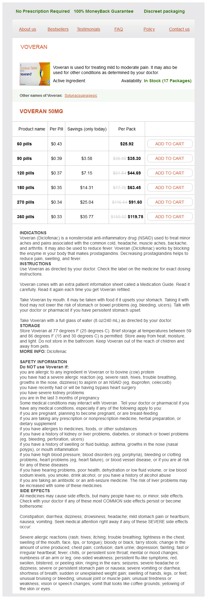
Voveran
| Contato
Página Inicial
"50 mg voveran generic visa, spasms in head".
Z. Spike, M.B.A., M.D.
Program Director, University of Kansas School of Medicine
Diseases
- Ocular convergence spasm
- Frontometaphyseal dysplasia
- Prothrombin deficiency
- Metacarpals 4 and 5 fusion
- Orofaciodigital syndrome Thurston type
- Hairy ears
- Large B-cell diffuse lymphoma
- Cardiomyopathy, fatal fetal, due to myocardial calcification
- Splenic flexure syndrome
They do spontaneously resolve over a interval of years but muscle relaxant names effective 50 mg voveran, if troublesome spasms hindi meaning generic 50 mg voveran visa, might require ablative therapy similar to laser or excision muscle relaxant vs pain killer voveran 50 mg generic amex. Varicosities Vulval varicosities are common throughout pregnancy and some thrombose spontaneously after delivery muscle relaxant injections voveran 50 mg discount on-line. They are often associated with varicosities on the decrease limbs, but when isolated to the genitalia the patient should be additional investigated to exclude an obstructive pel vic lesion. Disorders of lymphatics Acute lymphoedema Some swelling might occur in illnesses similar to candidiasis or acute eczema but resolves quickly with applicable remedy of the situation. The history is that of acute swelling, typically related to intercourse the place it might be strain induced. Type I hypersensitivity contact urticarial reactions to latex are an growing drawback. There is instant swelling of the labia after using latex condoms and it could additionally happen if healthcare staff put on latex gloves for examination. The vulva becomes thickened and indurated and may be more susceptible to attacks of cellulitis. Lymphangiectasia Small lymphatic vesicles (lymphangiectasia) could develop on a background of persistent lymphoedema. The lesions have a verrucose look and are sometimes incorrectly diagnosed as viral warts. Congenital lesions may have imaging research to establish if there are deeper lymphatic abnormalities. British Association of Dermatologists tips for the administration of lichen sclerosus, 2018. Vulvovaginal lichen planus remedy: a eleven 12 13 14 15 16 17 18 19 survey of present practices. Genital ulcers as initial manifestation of Epstein Barr virus an infection: two new cases and evaluation of the literature. It is therefore only examined in detail in circumstances where there are specific symptoms to immediate vaginal examination. The vagina consists of a nonkeratinized squamous epithelial lining supported by connective tissue and sur rounded by round and longitudinal muscle coats. The muscle is connected superiorly to the fibres of the uterine cervix, and inferiorly and laterally to the pubococcygeus, bulbospongiosus and perineum. The lower finish of the epithelium joins, close to the hymen, the mucosal compo nents of the vestibule and superiorly extends over the uterine cervix to the squamocolumnar junction. The vaginal epithelium has a longitudinal column within the ante rior and posterior wall, and from each column there are quite a few transverse ridges or rugae extending laterally on each side. The squamous epithelium through the reproductive years is thick and wealthy in glycogen. Vaginal an infection Between puberty and the menopause the vaginal lacto bacilli keep a pH between three. Before puberty and after the menopause, the upper pH level and urinary and faecal contamination enhance the risks of an infection. Vaginal atrophy can also happen within the postpartum interval with the hypooestrogenic state throughout lactation. Normal physiological vaginal discharge consists of a transudate from the vaginal wall, squames containing glycogen, polymorphs, lactobacilli, cervical mucus and residual menstrual fluid, in addition to a contribution from the greater and lesser vestibular glands. Vaginal discharge varies in accordance with oestrogen ranges through the menstrual cycle and is a traditional physiological prevalence. Nonspecific vaginitis may be related to sexual trauma, allergy to deodorants or contraceptives, and chemical irritation from topical antimicrobial remedy. Nonspecific an infection may be further provoked by the presence of international bodies, for instance ring pessary, continual use of tampons and the presence of an intrauterine contracep tive device. Bacterial vaginosis Bacterial vaginosis has been beforehand related to the organism Gardnerella vaginalis but a variety of organisms, together with Mobiluncus spp. Examination will reveal a thin grey�white discharge and a vaginal pH elevated to larger than 5. The prognosis can also be confirmed by including a drop of vaginal discharge to saline on a glass slide and adding one drop of 10% potassium hydroxide. Corynebacterium Mycoplasma Candida albicans + + - - - - - - - - - - - - - - - + + + - - + + + + - + + - - - - - - + + - - - - + - - +. Bacterial vagi nosis could also be associated with elevated danger of preterm labour [2], pelvic inflammatory disease and postopera tive pelvic an infection [3,4]. The therapy of bacterial vaginosis is with metronidazole, both as 200 mg 3 times a day for 7 days or as a single 2g dose. Trichomoniasis Trichomoniasis is a sexually transmitted illness brought on by the parasite Trichomonas vaginalis. Symptoms usually seem 5�28 days after publicity and include a yellow� green vaginal discharge, often foamy, with a robust odour, dyspareunia and vaginal irritation. Treatment is with metronidazole 400 mg thrice day by day for 7 days or tinidazole 2 mg as a single dose. As this may be a sexually transmitted disease, diagnosis ought to prompt the gynaecologist to refer the patient to a genito urinary drugs clinic for contact tracing. It is brought on by any of the species of Candida, of which Candida albicans is the most common. This is an an infection that causes vaginal irritation and vaginitis, which ends up in itching, burning, soreness and a traditional whitish or whitishgrey cottage cheeselike discharge. The irritation and irritation spreads throughout the vulva and can also contain the perianal pores and skin. Candida could be transmitted to a sexual associate, in whom it might possibly cause red patchy sores near the pinnacle of the penis or on the fore skin, inflicting a extreme itching and burning sensation. Candida albicans usually causes an infection when produc tion of lactic acid by lactobacilli is disturbed, resulting in a change in the pH in the vagina and subsequent over growth of Candida. Diabetics and patients using antibi otics for different infections have an elevated incidence of candidiasis. Candida is incessantly found within the vagina however it could also be a half of the intestinal flora. The analysis is usually made on inspection, however a swab from the contaminated area will confirm the prognosis in culture. Treatment for vaginal candidiasis is primarily with antifungal pessaries or cream inserted excessive into the vagina. Singledose prep arations provide the advantage of compliance, and imida zole drugs (clotrimazole, econazole and miconazole) are efficient in short programs of 1�14 days based on the preparation. Oral treatment can be obtainable within the type of fluconazole or itraconazole and these remedies are usually extremely efficient at eradicating the disease. Some 10% of ladies who contract candidiasis will develop recurrent illness � that is notably doubtless if there are predisposing elements, corresponding to being pregnant, diabe tes or oral contraceptive use. If bacteriological affirmation of recurrent disease is made, numerous longterm deal with ments could be prescribed. These embody fluconazole 100 mg orally every week for 6 months, clotrimazole 500mg pessary weekly for 6 months or itraconazole Benign Diseases of the Vagina, Cervix and Ovary 813 400 mg every month for 6 months. Recurrent vaginal candidiasis wants systemic investigation prior to longterm treatment. There are numerous surgical options, including incision and remedy with silver nitrate, marsupialization (which entails extensive excision, drainage and eversion of the cyst mucosa to the vaginal skin), fistulization (inci sion, drainage and insertion of a catheter for 2�4 weeks), carbon dioxide laser incision and drainage, and needle aspiration of cysts. However, unusual vaginal lesions should elevate suspicion, notably if the affected person or companion has just lately trav elled overseas. There is normally a single, painless, properly demarcated ulcer with indurated edges, related to lymphadenopathy. Secondary lesions embrace condylo mata lata, mucous patches and snailtrack ulcers. Vaginal atrophy this is seen not solely following the menopause, but also prior to puberty and during lactation. Examination exhibits loss of rugal folds and prominent subepithelial vessels, generally with adjacent ecchymoses.
Buckels (Cowslip). Voveran.
- What other names is Cowslip known by?
- How does Cowslip work?
- Are there safety concerns?
- Inflamed nasal passages or sinusitis when taken with gentian root, European elder flower, verbena, and cowslip flower (SinuComp, Sinupret).
- Dosing considerations for Cowslip.
- What is Cowslip?
Source: http://www.rxlist.com/script/main/art.asp?articlekey=96202
The genetic defects underlying DiGeorge syndrome have been elusive muscle relaxant list 50 mg voveran discount with mastercard, but knowledge is advancing spasms lower back discount voveran 50 mg online. A deletion of up to gut spasms order voveran 50 mg visa 3 megabases in chromosome 22 (22q11) has long been identified to be related to DiGeorge muscle relaxant 4211 voveran 50 mg order, based mostly on kindred studies in families with the syndrome. In people, the defect is autosomal dominant, with variable penetration, meaning that one copy of the gene deletion is enough to cause illness and that the illness severity various from family member to member of the family. A comparable deletion generated experimentally in mice yielded a model of the cardiovascular options of DiGeorge syndrome (Lindsay et al, 1999). The proper dorsal and left ventral cushions are throughout the conus area and are essential for pulmonary and aortic valve formation, and for completion of septation between the ventricles at the degree of the pulmonary and aortic valves. To align the septated great arteries immediately over the ventricles, the arterial trunk should shift to the left. Anatomically, the normal conus is a muscular "neck" inside the best ventricle between the tricuspid and pulmonary valves. Heterozygous mutants, which are analogous to the human disease state, showed more variable illness. The cardiac defects primarily affected the fourth pharyngeal arch and triggered abnormal patterning of the nice arteries in 50% of embryos in one research (Lindsay et al, 2001). This group had 1 heterozygous mouse mutant out of 14 that additionally exhibited parathyroid and thymic insufficiency. Possibly, people are extra delicate to gene dosage and exhibit the total range of defects in the haploinsufficient state. There could additionally be different modifying genes as nicely, because there appears to be an important contribution of genetic background to the phenotype in mice, with more extreme arch anomalies presenting in more inbred strains (Jerome and Papaioannou, 2001). This is a confounding issue of many gene focusing on methods and maybe a clue as to why significant phenotypic variation can occur in people with equivalent mutations. Further along the outflow tract, the pulmonary artery and aorta are extra left-right to each other, because the spiral extends. This rotation entails not only the truncal cushions but the rotation of the myocardial tube as well (Bajolle et al, 2006; Lomonico et al, 1986). The left-right orientation of the valves is mostly preserved (aortic rightward), though can range within a 90-degree range from instantly anterior-posterior to immediately aspect by facet. L-transposition, aortic leftward, is most frequently related to L-looped ventricles or heterotaxy syndromes and is rare in isolated transposition. The views above are coronal sections via the pulmonary trunk, considered from the ventral facet. The embryo under is shown from a transverse section via the pulmonary trunk, revealing the pulmonary valve leaflets. The valves undergo intensive remodeling of the extracellular matrix; in the case of the semilunar valves, this results in a fibrous layer (primarily collagen) on the arterial aspect, a spongiosa layer (glycosaminoglycans) within the center, and an elastic layer (elastin) on the ventricular facet (Combs and Yutzey, 2009; Hinton et al, 2006; Shelton and Yutzey, 2007). In the case of the semilunar valves, this reworking strategy of the endocardial cushions continues by way of the last trimester of pregnancy and into the neonatal interval (Aikawa et al, 2006; Hinton et al, 2006). There is controversy as to how the septum extends so shortly to type a wall between the left and right ventricles. Part of the reply seems to be speedy proliferation of these myocytes, which retain the power to divide whilst a working myocardium. This property is misplaced soon after delivery, as the mature myocardium is for essentially the most part incapable of proliferation. Nearly 90% of those small defects had been closed by 10 months of age (Roguin et al, 1995). These information and others recommend continued lowlevel proliferation in human ventricles after delivery. We have said that as the muscular septum grows and extends from the ventricular apex, the separation between the left and proper ventricles turns into nearly complete. There are two essential regions of the whole ventricular septum which are completed by endocardial cushions, as discussed within the previous sections. An necessary feature of this course of not mentioned earlier is the reality that these regions of septum comprise muscle in addition to cushion tissue. These migrating myocytes are nonproliferative, and the whole inside curvature is involved. Thus, the mitral-aortic continuity discussed earlier is completely fibrous and devoid of muscle. The left and right ventricles differ not solely by their relative positions to each other, but also of their primary muscular structure and in their perform. In the embryo, the myocardium thickens and arranges itself in processes generally recognized as trabeculation and compaction. Trabeculation refers to projections of muscular tissue into the lumen of the ventricle, such that the internal surface is not smooth-walled, but ridged. Orientation as shown by the arrows: P (posterior), A (anterior), R (right), and L (left). Here the atrial septum primum (*) is fused with the endocardial cushions inferiorly; not seen is the foramen ovale that enables steady rightto-left atrial shunting within the fetus. The arrowhead reveals a small residual ventricular septal defect in the inlet (posterior) septum, not yet closed by endocardial cushion tissue. Soon afterward, a model new crescent of atrial septum varieties on the proper atrial facet of the septum primum and begins extending alongside the septum primum. During fetal life, blood flow from the best atrium into the left prevents fusion of those two septae. After birth and separation from the low-resistance placenta, the left atrial stress rises considerably and the atrial shunt reverses. This causes the septum primum to impact a seal towards the septum secundum, often leaving a small opening often known as the foramen ovale. A patent foramen ovale, usually three mm or less with a trivial degree of left-to-right circulate, is a standard discovering in a new child and even in lots of older youngsters and adults. Failure of the cushions to kind the inferiormost part of the atrial septum leads to a primum atrial septal defect. The vitelline veins return from the yolk sac, a construction that communicates with the primitive gut through the vitelline duct. Distally, the vitelline veins regress together with the duct and proximally lose their connection to the sinus venosus by 6 weeks. As the liver develops, the umbilical veins develop connections to the liver venous plexus, and the connection to the sinus venosus involutes. This flow is primarily directed toward the atrial septum, and therefore primarily through the foramen ovale into the left atrium and systemic circulation. The left and proper widespread cardinal veins are comparatively short segments that connect with the sinus venosus. On both sides, anterior and posterior cardinal veins be a part of to kind the frequent cardinal vein, the anterior carrying the venous return from the higher physique and the posterior from the lower body. The right anterior and proper widespread cardinal vein will type the superior vena cava within the mature circulation. The left frequent cardinal vein and left phase of the sinus venosus form the coronary sinus, which receives venous return from the coronary system. The pulmonary veins grow in progressively from the creating lung vasculature, first as a typical pulmonary vein from each lungs. It is along these arches that the neural crest migrates into the conotruncus and helps growth of the arch arteries. Again these are paired structures, and there are a complete of six pairs, although not all are patent at the same time. Stages of systemic vein growth are shown in A to D, during which the paired umbilical, vitelline, and cardinal veins give rise to the only umbilical vein, liver vasculature including the portal system, and the inferior and superior vena cavae. Stages of pulmonary vein development are proven by week gestation as labeled; the frequent pulmonary vein progressively absorbs into the back wall of the left atrium. The left limb of arch four stays in continuity with the aortic root, collectively making up the true aortic arch and its first department, the brachiocephalic (innominate) artery. The proper limb of arch 4 turns into a half of the best subclavian artery, which retains its proximal connection with the brachiocephalic artery (Waldo et al, 1996). In some circumstances, either independently or as part of another developmental cardiac defect, the right limb of arch four remains patent and the left forms the left subclavian artery-this varieties a right aortic arch with mirror-image branching.
Syndromes
- Bruising
- Not sleeping very well
- An overwhelming urge to make the movement
- Being around cigarette smoke while in the womb or after being born
- Other infections
- Eye care products
- Excessive bleeding