

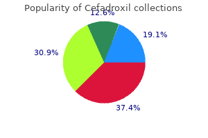
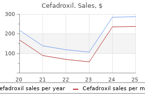
Cefadroxil
| Contato
Página Inicial
Achilles J. Pappano PhD
- Professor Emeritus, Department of Cell Biology and Calhoun Cardiology Center
- University of Connecticut Health Center, Farmington
https://health.uconn.edu/cell-biology/faculty-and-staff/achilles-j-pappano/
The corneal endothelial cells diminish in number significantly faster than is normal for aging antibiotic 1p 272 purchase 250 mg cefadroxil with amex, ultimately leading to chronic edema of the cornea antibiotics news purchase cefadroxil 250 mg with amex. The endothelium may then decompensate antibiotics for dogs amoxicillin discount cefadroxil master card, either immediately if the residual endothelial cells are too few antibiotics you can give a cat buy cefadroxil cheap, or sometime later virus like chicken pox discount 250 mg cefadroxil fast delivery, after the endothelial cell population declines further with age. Although cataract surgery is the most common intraocular procedure performed, bullous keratopathy may similarly occur following other intraocular procedures, for example, multiple glaucoma surgeries or retinal surgeries. Keratoconus is a condition generally presenting in teenagers and young adults in which the cornea is more ectatic. Both genetic and environmentaVacquired factors may be involved, since keratoconus is associated with both hereditary connective tissue disorders such as Marfan syndrome, as well as with excessive eye rubbing such as in chronic atopic conjunctivitis or chronic blepharitis associated with Down syndrome. Corneal buttons with ulcerative keratitis may or may not reveal the offending microorganism when special stains are performed. Lack of microorganisms on histology despite a history of positive cultures is generally attributable to antecedent anti-microbial therapy, as an attempt is generally made to sterilize the ulcer prior to keratoplasty. Residual suture material may be present, or may be absent if all the sutures were removed at some point after the initial procedure. The most essential histologic feature of graft failure is generally endothelial cell loss. The antecedent event can range from previous accidental trauma or previous surgical trauma to specific disease entities such as retinal vascular disease (including diabetes, or central retinal artery or vein occlusion), intraocular infection (endophthalmitis), chronic inflammatory disease, or primary retinal detachment. Surgical methods of removing an eye include enucleation, in which the entire eye is removed intact, or evisceration, in which only the intraocular contents (lens, retina, uvea) and possibly cornea are removed with retention of the sclera in the eye socket to house the orbital implant. Exenteration, in which the eye along with surrounding orbital soft tissues are removed, is sometimes required to achieve complete excision of an eyelid/orbital tumor (see later). The key histopathologic finding in most cases of secondary glaucoma is an anterior chamber angle closed by peripheral anterior synechiae. The term "closed angle" means that the anatomical angle normally formed by the cornea and iris, and occupied by the trabecular meshwork (the main outflow channel for aqueous humor), is occluded by the peripheral iris. These adhesions may be induced by chronic inflammation, such as in various forms of chronic uveitis, or by the abnormal proliferation of capillaries (neovascular glaucoma), as seen in proliferative diabetic retinopathy or retinal vascular occlusion (see later). Occasionally, in contrast to the closedangle appearance, post-contusion angle recession may be seen in cases of blunt trauma. Retinal detachment may be idiopathic, trauma induced, related to inflammation or infection, or secondary to proliferative retinopathy of various etiologies (diabetic, post-vascular occlusion, and so on). In cases of old central retinal vein or artery occlusion, the retina exhibits atrophy and cystoid degeneration of the inner two-thirds of the retina, lipoproteinaceous exudates, and in the case of venous occlusion, often a persistence of hemorrhage. There may also be a neovascular epiretinal membrane with tractional retinal detachment and vitreous hemorrhage. As with neovascular glaucoma of any etiology, eyes with severe diabetic retinopathy or central retinal vascular occlusion also exhibit atrophy and possibly cupping of the optic nerve. Endophthalmitis is generally treated with intravitreal antibiotics and/or antifungals, and possibly vitrectomy surgery to clear the abscess. However, in severe cases treatment may not be successful, and evisceration or enucleation may ultimately be performed. Often there is loss of intraocular contents such as the lens and a portion of the uvea, and possibly also the retina. If an attempt has been made by the surgeon to salvage the ruptured globe, but the eye later has to be enucleated because it has become blind and painful, the histopathologic findings vary depending on the degree of chronicity; in cases of recent rupture, the findings may be the same as described above. In cases with a more distant history of rupture, there may be total retinal detachment with gliosis and loss of intraocular contents, intraocular granulation tissue or fibroconnective tissue emanating from the rupture site, and often osseous metaplasia of the retinal pigment epithelium (see later). Angle closure or post-contusion angle recession may also be present depending on the location of the rupture and the nature of the traumatic forces. When an eye has undergone a previous insult such as severe trauma, complications of surgery, proliferative retinopathy, or chronic inflammation, and has a chronic total retinal detachment, a gradual degenerative process eventually ensues in which the eye undergoes shrinkage, that is, phthisis (from the Greek verb "to wither"). Clinically, the eye has hypotony (very low intraocular pressure) and is visibly shrunken. Histologically, the retina is totally detached, with marked diffuse atrophy and architectural distortion of all layers of the eye including such severe retinal gliosis as to render the retina barely recognizable. Gross room processing of enucleated globes follows a standard protocol to generate the pupil-optic nerve (p. The figure shows the standard transverse plane that is often used for cutting the section, but the globe may be opened along any plane that best captures the particular area of interest. For eyes enucleated due to intraocular tumors, especially primary uveal melanoma, the tumor may be localized by transillumination prior to cutting the eye. Transillumination is achieved by shining onto the cornea a very bright light source that is about 1 em or less in diameter. The normal uvea will not block the transmission of light, and hence the sclera will glow. However, a uveal tumor will block light transmission, thereby casting a shadow over the corresponding portion of sclera. The superior cap (also known as the superior calotre) and inferior cap are usually not processed unless abnormalities are seen or the eye has an intraocular tumor. The most common primary intraocular neoplasm is melanoma of the uvea (choroid, ciliary body, or less commonly, iris). Melanomas of the iris have a better prognosis than do melanomas of the ciliary body and/or choroid. Although some intraocular melanomas can be treated by excision or brachytherapy, some patients do not seek medical attention until the melanoma has become so large that the only course of management is enucleation of the globe. Other factors associated with a worse prognosis include necrosis and mitotic figures. The usual age for clinical presentation in nonfamilial cases is 1 year, whereas familial cases are generally screened within the first few weeks of life and therefore present earlier. In nonfamilial cases, the tumor is generally large and unilateral and accordingly treated with enucleation. The tumor may be much larger in one eye than the other, in which case the eye with the large tumor is enucleated, whereas the eye with smaller tumor(s) may be salvaged via laser or cryotherapy applied to the tumor foci, possibly with antecedent adjuvant systemic chemotherapy. A few centers utilize intraarterial chemotherapy (delivered to the ophthalmic artery) to treat unilateral or bilateral tumors, thereby potentially salvaging eyes that would otherwise need to be enucleated. Cytologically, the tumor is composed of cells with hyperchromatic, oval-shaped nuclei with scant cytoplasm; the nuclei are densely packed together and thus show nuclear molding. If optic nerve invasion past the lamina cribrosa or massive choroidal invasion is seen on histology, then the patient is treated with adjuvant systemic chemotherapy. Infiltrative lesions include dacryoadenitis, sarcoidosis, lymphoid hyperplasia, and lymphoma. The salivary glands are exocrine organs that secrete components of saliva to both break down carbohydrates and lubricate the passage of food. There are three major paired salivary glands: the parotid, the submandibular, and the sublingual. The parotid glands are the largest major salivary glands and are located between the ramus of the mandible and the mastoid process. Each gland is composed of a superficial lobe and a smaller deep lobe, and the facial nerve is intimately associated with both lobes. The submandibular glands (also referred to as the submaxillary glands) are located just medial to the body of the mandible, and are smaller than the parotid glands. The sublingual glands are the smallest of the major salivary glands and are located in the floor of the mouth between the genioglossus muscle and the mandible. The major salivary glands are enclosed by a connective tissue capsule and divided into lobules composed of ducts and acini, whereas the minor salivary glands are unencapsulated. The acini are composed of mucinous and/or serous epithelial cells surrounded by a layer of myoepithelial cells which contract to aid in the movement of glandular secretions. Mucous cells are more rounded, have basally oriented nuclei, and contain abundant clear mucin. Intercalated ducts are also the source of reserve cells that can repopulate the acinar system. The intercalated ducts join to form striated ducts, which merge to form interlobular ducts, which ultimately empty into the large named ducts. Biopsies of many salivary gland lesions are taken prior to surgery to characterize the lesion and direct management. Core needle and incisional biopsies of the parotid gland can damage the facial nerve branches so are rarely performed. Some salivary gland masses are removed in their entirety without a previous tissue diagnosis. Superficial parotidectomy remains the initial procedure of choice for benign parotid gland tumors. Submandibular gland resections are usually performed without taking any significant periglandular soft tissue, although more tissue may be included with the specimen for submandibular or parotid tumors that are known or suspected to be malignant. The rare sublingual gland tumors necessitate a resection of the floor of mouth that is qualitatively similar to those performed for mucosal-based squamous carcinomas of the same area. In general, salivary gland specimens are small enough to prosect the day of receipt. The deep (covered by muscle and fascia) and superficial (covered by subcutaneous fat) surfaces of the parotid gland can sometimes be discerned, and should be differentially inked. The specimen should be serially sectioned, and any mass or focal lesion described including dimensions, color, texture, and distance to the margins. Four to five sections of the tumor, including representative areas of the closest inked margins, should be taken. For the parotid gland, the surrounding gland and any periglandular soft tissue should be thoroughly searched for lymph nodes, which should be separately submitted. Sialadenitis, or inflammation of the salivary glands, can be divided into bacterial causes, viral causes, and autoimmune disease. Bacterial sialadenitis is rare and is generally a result of obstruction by stones (sialolithiasis). Microscopic sections can show abscesses, cystic degeneration, and/or bacterial colonies. Viral sialadenitis is most commonly caused by mumps (paramyxovirus), but can also be associated with Epstein-Barr, coxsackie, influenza A, and parainfluenza viruses. Grossly infected glands are boggy and edematous, and chronic inflammation may be seen microscopically. On physical examination, bilateral, symmetric enlargement of the salivary and lacrimal glands is seen. Diagnosis may be facilitated by a minor salivary gland biopsy, typically taken from the inner lip. Microscopically, in early disease, there is a lymphoplasmacytic septal inflammatory infiltrate with little to no abnormality of the parenchyma. The so-called "focus score," a nodular collection of >50 lymphocytes, is considered diagnostic of autoimmune sialadenitis in the appropriate clinical context. In late disease, acini may be completely absent leaving only isolated residual ducts, termed "epimyoepithelial islands," with an associated dense intraepitheliallymphocytosis. Chronic sialadenitis (or chronic sclerosing sialadenitis) is usually unilateral and clinically can mimic a true salivary gland neoplasm. Chronic sialadenitis most commonly results from sialolithiasis with obstruction, although some cases may be due to radiation therapy or duct strictures. It can have no structural or obvious etiology and recently most of these cases have been shown to be an autoimmune-related lgG4-related sclerosing disease similar to that occurring in the pancreas. Histologic examination of the gland early in the disease process shows dilated ducts filled with secretions with an associated lymphocyte and plasma cell-rich infiltrate, occasionally with germinal center formation. Surgical excision may be required, either to definitively rule out a neoplasm or just for symptom relief. Necrotizing sialometaplasia is a rare inflammatory/destructive lesion that simulates malignancy and is thought to occur because of ischemic injury. Clinically, the lesion consists of a sharply defined and deep ulcer that develops rapidly (over a few days) and can persist for months. Grossly, the lesion consists of loose tissue with a surface ulcer and no distinguishing mass lesion. Microscopically, the most typical feature is coagulative necrosis of the minor salivary gland lobules with a prominent associated inflammatory response. The lack of peripheral infiltration and the retained lobular architecture are keys to recognizing the lesion as benign. Patients present with unilateral or bilateral, painless, slowly enlarging parotid masses. They sometimes have associated cervical lymphadenopathy and/or nasopharyngeal swelling. Grossly, these lesions have multiple cystic spaces usually containing serous fluid. Below the epithelium, the cyst wall has dense lymphoid tissue with germinal centers. Occasionally, lesions within the gland can lack cystic change and appear similar to epimyoepithelial islands. Patients usually have bilateral disease by radiologic examination, even if there are no symptoms and no clinical mass in the contralateral gland. If the diagnosis is established by radiologic imaging and cytology, surgery is not necessary for other than cosmetic reasons. Benign lymphoepithelial cysts are unifocal lesions that occur in patients in their fifth and sixth decades. They most commonly involve the parotid gland, but are occasionally seen in the oral cavity. Grossly, they consist of well-circumscribed unilocular cysts with contents ranging from serous to mucoid to caseous; keratinous debris may be present.

Also termed nonspecific thyroiditis or focal autoimmune thyroiditis infections after surgery buy cefadroxil 250 mg visa, this is a common disorder usually discovered incidentally in surgically excised thyroids antibiotics used uti purchase cefadroxil 250 mg otc, and is found in 25% to 60% of patients in autopsy studies antibiotic 3 day generic cefadroxil 250 mg on line, and is most common in older women bacteria kingdoms discount cefadroxil 250 mg online. The lymphocytes can infiltrate follicles antibiotic resistance prevention purchase cheap cefadroxil, but there is no significant destruction or damage. Hashimoto thyroiditis, or chronic lymphocytic thyroiditis, is an autoimmune disease with thyroid enlargement and circulating antithyroid antibodies. There is a familial association, and Hashimoto thyroiditis is often associated with other autoimmune diseases. Patients can present with a hyperthyroidism or hypothyroidism and are typically in their sixth decade. Grossly, the thyroid is diffusely enlarged and firm with a mildly nodular surface. On sectioning, lobulation is accentuated due to fibrosis, and the gland is tan-yellow or off-white rather than the usual brown due to Chapter 24 · Thyroid I 407 the abundant lymphoid tissue. Microscopically, there are sheets of lymphocytes and plasma cells with abundant germinal centers. Florid inflammation often renders the residual follicles and follicular epithelium inconspicuous. The inflammatory infiltrate may extend into the perithyroidal soft tissue causing adherence of the gland at the time of surgery. Finally, there may be fibrous septa between the lobules, and the follicular epithelium may develop squamous metaplasia. A number of variants of Hashimoto thyroiditis occur, including fibrous (sclerosing), fibrous atrophy, juvenile, and cystic forms. Both of the fibrous variants show the same histologic findings, namely retention of a lobulated pattern with extensive and severe fibrosis. Atrophic follicular cells are more scattered but show Hiirthle cell change, and there is abundant chronic inflammation with germinal centers. Both of the latter variant forms are associated with marked hypothyroidism and high titers of antithyroid antibodies. The differential diagnosis for Hashimoto thyroiditis includes Riedel thyroiditis and lymphoma. The fibrous variant of Hashimoto thyroiditis may simulate Riedel thyroiditis with an enlarged fibrotic gland. However, the fibrosis is typically limited to the gland itself, whereas in Riedel thyroiditis there is severe adherence of the fibrotic gland to the neck soft tissues. The dense inflammation in Hashimoto thyroiditis can simulate lymphoma, and most thyroid lymphomas do arise in the setting of Hashimoto thyroiditis. The distinction lies in the absence of sheets of atypical lymphocytes, the lack of a strikingly prominent lymphoepithelial pattern, and the lack of clonality of the lymphocytes by flow cytometry or molecular studies. The issue of whether there is an increased risk of papillary carcinoma in Hashimoto thyroiditis is a controversial topic. Whether this is the case or not, the nuclear features of the epithelium can simulate those of papillary carcinoma (including nuclear crowding, clear chromatin, and occasional nuclear grooves) and develop frequently in the inflamed follicular epithelium. A true carcinoma arising in Hashimoto thyroiditis should stand out sharply from the neighboring follicular epithelium, as in uninflamed thyroid. Graves disease is also known as diffuse hyperplasia and is an autoimmune condition resulting in excess thyroid hormone production. It causes the majority of cases of spontaneous hyperthyroidism, occurs most often in the third and fourth decades, and is 5 to 10 times more common in women. Patients also develop a characteristic infiltrative ophthalmopathy with proptosis. Posttreatment or after long-standing disease, the gland may be somewhat nodular or fibrotic. Microscopically, the gland typically shows a low-power lobular accentuation due to increased septal fibrous tissue. Inflammation varies from none, to patchy lymphocytic, to lymphocytic with formation of germinal centers. The follicular epithelium has increased amounts of cytoplasm, is convoluted and irregular, and often assumes an almost papillary appearance. Treated cases have a variable morphology and often have cellular nodules mimicking adenomas. The differential diagnosis includes papillary carcinoma when the stellate outlines of follicles resemble papillae. The lack of cytologic changes of papillary carcinoma and the diffuse gland involvement are keys to the correct diagnosis of Graves disease. A peculiar form of fibrosing disease, Riedel thyroiditis is a chronic thyroiditis of unknown etiology which is more common in women, occurs most commonly in the fifth decade, and is commonly associated with fibrosing disease at other sites such as the mediastinum, retroperitoneum, and lung. Patients present with firm thyroid enlargement and local symptoms such as dysphagia, stridor, or dyspnea. Recurrent laryngeal nerve or sympathetic trunk involvement can lead to hoarseness or Horner syndrome; compression of the large vessels can lead to superior vena cava syndrome. Most patients are euthyroid at presentation, but many subsequently develop hypothyroidism. Grossly, the thyroid gland is usually received in irregular pieces because the fibrosis makes it difficult to remove surgically. Microscopically, the characteristic finding is dense and hypocellular eosinophilic fibrous tissue with scattered and patchy aggregates of lymphocytes, plasma cells, neutrophils, and eosinophils, without germinal centers or granulomas. Rare entrapped and atrophic thyroid follicles are seen without Hiirthle cell change. A characteristic finding is small veins with infiltrating lymphocytes and myxoid intimal thickening (features of occlusive vasculitis). The differential diagnosis includes hypocellular anaplastic thyroid carcinoma and fibrous or sclerosing Hashimoto thyroiditis. The lack of Hiirthle cell change and germinal centers, and the profound degree of perithyroidal fibrosis, rules out fibrous Hashimoto disease. Nodular hyperplasia (clinically termed multinodular goiter) is an extremely common disorder of the thyroid gland. With supplementation of iodine in the diet, nodular hyperplasia has also been related either to excess iodine intake with impaired organification or to genetic factors. It is much more common in women than men and typically presents in middle age as asymptomatic enlargement. Large goiters, however, can cause dysphagia, hoarseness, or stridor and can extend into the upper mediastinum. A small percentage of patients present with hyperthyroidism (toxic multinodular goiter). On sectioning, the nodules may be semitranslucent, glistening, fleshy, red-brown, tan, and solid or, more commonly, show varying degrees of degeneration with cystic change, hemorrhage, fibrosis, and calcification. The nodules may be quite cellular with tightly packed follicular epithelium and little colloid, resembling follicular adenomas or carcinomas. Around cystic areas there is often fibrosis with variably sized foci of dystrophic calcification and Cllojbr:u · n. Follicular carcinomas are almost always unifocal and have tan to brown, solid cut surfaces. Microscopically, the growth pattern ranges from well-formed follicles throughout, to solid, to trabecular. The oncocytic variant (Hiirthle cell) is the main histopathologic variant and compromises 20% to 25% of all follicular carcinomas. To be classified as oncocytic variant, a tumor should be composed of at least 75% oncocytic cells. The distinction of minimally invasive follicular carcinoma from adenoma lies in capsular and/or vascular penetration alone (Table 24. Strict adherence to these criteria is necessary to assure that the diagnosis of carcinoma is correct. Finally, poorly differentiated (insular) carcinomas can be similar to widely invasive follicular carcinomas, but the former have cells with less cytoplasm, minimal follicular architecture, marked mitotic activity, and necrosis. In general, minimally invasive follicular carcinomas have an excellent prognosis with an approximately 15% long-term mortality. There is evidence that the presence and extent of vascular invasion may provide further prognostic information for patients with minimally invasive follicular carcinomas. Carcinomas that lack vascular invasion have a better outcome (<5% long-term mortality), while the presence of four or more foci of vascular invasion has been shown to correlate with increased tumor-related mortality. Widely invasive follicular carcinomas have a poorer long-term mortality approaching 50%. Because the classification of poorly differentiated carcinoma is somewhat controversial, epidemiologic data are difficult to obtain. In general, however, it represents much <5% of thyroid carcinomas in the United States, but between 4% and 7% in Italy and some Latin American countries. Most cases present as sizable asymptomatic masses, with or without pathologically enlarged regional lymph nodes. There is occasionally a history of a long-standing mass with recent, rapid growth. It is not uncommon for well-formed capsules to be present around some of the nodules. The terminology has become somewhat confusing because papillary architecture, although very commonly present, is not necessary for diagnosis. There is a close link with previous radiation exposure, particularly in younger patients. Cystic change is relatively common, particularly in lymph node metastases, and the bulk of nodal metastatic disease may far outstrip the volume of tumor in the thyroid gland. Architectural patterns include papillary, trabeculat; micro- and macrofolliculat; solid, and cystic. In tumors with follicles, the colloid is typically described as "bright" or dark red relative to the normal thyroid. Distinct nuclear features include marked crowding with overlapping of adjacent nuclei, pale Cllojbr:u. Microscopically, it is typically composed of a variable admixture of spindle cells, epithelioid cells, and giant cells. There is often geographic necrosis, and typically the remaining thyroid gland is obliterated by tumot: On thorough examination, a residual, well-differentiated carcinoma, either papillary or folliculat; is sometimes identified. Immunohistochemistry for cytokeratin is positive in approximately 80% of cases, and for epithelial membrane antigen in 30% to 50%. These stains are useful for diagnosis of tumors in which no obvious carcinomatous differentiation is present on H&E in order to confirm that the neoplasm is a carcinoma rather than a high-grade sarcoma. C-cells secrete calcitonin, a peptide that causes increased renal excretion of calcium and inhibits osteoclasts to prevent calcium liberation from bone. Sporadic tumors are solitary, whereas familial tumors are often multifocal and bilateral. The typical features are sheets, nests, or trabeculae of polygonal, round to oval cells with moderate to generous amounts of eosinophilic to amphophilic cytoplasm. The nuclei are round to oval with a coarse and somewhat granular neuroendocrine chromatin. Reactive, or nonneoplastic, C-cell hyperplasia occurs in aging and a number of other thyroid diseases, most notably hyperparathyroidism and lymphocytic thyroiditis. Although the histology of C-cell hyperplasia is Chapter 24 · Thyroid I 41 7 somewhat controversial, a few generalities apply. First, reactive C-cell hyperplasia is usually unilateral and is not identifiable by H&E examination alone. Second, if there are aggregates of C cells >50 in number, in nodules, bilaterally, or diffusely, neoplastic C-cell hyperplasia is diagnosed. Neoplastic C-cell hyperplasia progresses to medullary carcinoma when C-cells extend through the follicular basement membrane into the stroma. Helpful histologic features of microcarcinoma include: (i) nuclear pleomorphism, (ii) expansile growth pattern with C-cell clusters spilling out of follicles, (iii) sclerotic stroma, and (iv) amyloid deposition. Patients who do not have metastatic disease are usually cured by total thyroidectomy, and their 10-year survival approaches 100%. Lymph node metastases are very common, and common distant metastatic sites include the lungs, liver, and bone. The overall10-year survival for patients with cervical lymph node metastases is approximately 70% to 80%, and for patients with distant metastases 40% to 50%. Patients with papillary carcinoma that has not metastasized distantly have an excellent prognosis, particularly those younger than 45 years of age, in whom survival approaches baseline. The sensitivity for the detection of lesions requiring surgical management is up to 98%, but specificity lags. The recent introduction of the Bethesda System for Reporting Thyroid Cytopathology (Am] Clin Pathol. Exceptions include paucicellular samples with marked cytologic atypia; solid nodules with inflammation. If only macrophages and noncolloidal cyst fluid are present, and there is a solid clinical/sonographic lesion, the assumption should be made that the lesion has not been adequately sampled. In more reassuring clinical/sonographic cases, cyst fluid only may be consistent with a benign process. It should be clear that "cyst fluid only" requires careful contextual interpretation. This is a category with high negative predictive value (0% to 3% malignancy rate on follow-up). Smears generally show significant amounts of colloid and variable degrees of cellularity. Benign follicular nodule is the most common pathologic correlate, but the presence of inflammatory cells or fibrous stroma may indicate a thyroiditis. Benign follicular nodules, the most common diagnosis in thyroid aspirates, are characterized by benign follicular cells and colloid in variable proportions, variably sized follicular groups, Hi.
Grossly virus games order 250 mg cefadroxil overnight delivery, they are polypoid or pedunculated antibiotic 93 cefadroxil 250 mg purchase, pink to red antibiotics korean 250 mg cefadroxil with mastercard, and have a smooth surface bacterial meningitis symptoms discount cefadroxil 250 mg fast delivery. Pregnancy-related lesions often regress postpartum and thus may be hormonally driven bacteria ua rare purchase cefadroxil 250 mg line. Hemangioma and lymphangioma are benign tumors composed of abundant blood or lymphatic vessels, respectively. Microscopically, they consist of blood vessels ranging from small capillaries to large cavernous spaces. The endothelial lining cells can be plump and may have mitotic activity, a feature more common in children. There are a number of named histologic variants, most of which have no clinical significance. About 75% occur in the head and neck and, when presenting in the oral cavity, they are almost always found in children younger than 3 years. Histologically, they typically consist of very dilated lymphatic channels lined by bland, inconspicuous endothelial cells, with intraluminal eosinophilic material, lymphocytes, and occasional red blood cells. Both hemangioma and lymphangioma are benign lesions cured by conservative excision. Large lymphangiomas are often de bulked, often via serial resections to avoid major morbidity. There are frequently associated collections of extravasated red blood cells and hemosiderin-laden macrophages. A characteristic feature that is sometimes seen is the pale, eosinophilic hyaline globule, which probably represents degenerating red blood cells. Granular cell tumors are benign, slow growing tumors of neural origin that occur at many anatomic sites. Approximately 50% occur in the head and neck region, and half of these occur in the tongue. They also occur in the buccal mucosa, floor of mouth, and palate, are twice as common in women as men, and approximately 10% to 20% are multiple. The nuclei are small, oval, and hyperchromatic with minimal atypia and no mitotic activity. Conservative excision is the treatment of choice, with a risk of recurrence of <10%. Malignant granular cell tumors (as covered in the Soft Tissue chapter) are very rare but do occur. Peripheral ossifying fibroma is a reactive proliferation of fibrous tissue on the gingiva which shows focal bone formation. They are essentially exclusive to the gingiva, particularly along the incisors, and present as sessile pink nodules, usually with surface ulceration. The lesions should be excised down to the periosteum but will recur in 15% to 20% of cases. Peripheral giant cell granuloma is another reactive proliferation of the gingiva, particularly along the incisors, caused by chronic irritation. It presents over a wide age range, particularly in middle-aged to older adults, as a solitary broad-based nodule that is reddish or blue and <2 em in diameter. The differential diagnosis includes brown tumor of hyperparathyroidism, cherubism, and central (intraosseous) giant cell granuloma. Congenital granular cell epulis is a rare benign mesenchymal tumor that classically arises from the anterior alveolar ridge of a newborn. Microscopically, it consists of sheets of large cells that have abundant, granular, eosinophilic cytoplasm and round to oval, bland nuclei. By immunohistochemistry, the cells are positive only for vimentin, and specifically are negative for S-100. Congenital granular cell epulis is not the newborn equivalent of a granular cell tumor and shows no neural differentiation. The tumor stops growing at birth and regresses over time, but most cases still require surgical resection. Staging is extremely important for clinical management and establishing prognosis. A specific and very important point in staging involves bone involvement by tumor. It is important to note that mucosal melanomas of the head and neck, including the oral cavity, have their own staging system. As with carcinomas at all upper aerodigestive tract sites, margin status, tumor differentiation, and the presence or absence of perineural or lymphovascular space invasion should be reported. Perineural invasion is particularly common in oral cavity carcinomas and is correlated with a poorer prognosis. The pattern of infiltration as well as the presence or absence of a host inflammatory response should also be reported, because both features have been correlated in many studies with a higher rate of local recurrence, a poorer prognosis, or both. Some recent studies have developed formal grading systems for these features that have promise for clinical implementation, particularly for early stage oral cavity sec, but these are not ready for active implementation. Depth of invasion, particularly for T1 and T2 tumors, although not reflected in the staging system specifically, is important for clinical management and prognosis, and so should be reported. Head and neck squamous cell cancer and the human papillomavirus: summary of a National Cancer Institute State of the Science Meeting, November 9-10,2008. Human papillomavirus-positive basaloid squamous cell carcinomas of the upper aerodigestive tract: A distinct clinicopathologic and molecular subtype of basaloid squamous cell carcinoma. The larynx is a unique organ designed to produce phonation by modulation of the respiratory airstream. It is composed of several cartilaginous structures: the thyroid, cricoid, and arytenoid cartilages and the epiglottis. The hyoid bone sits above and is connected to the larynx by the thyrohyoid membrane. The glottis includes the true vocal folds or cords, below, and the false folds or cords, above. The space between them is called the ventricle, and its deeper recess, the saccule. The cords are manipulated by muscles that attach to and move the arytenoid cartilages, which sit at the posterior aspect of the vocal folds. The larynx can be divided into three compartments for tumor management and staging purposes: the supraglottis, glottis, and subglottis. The supraglottis includes the epiglottis, aryepiglottic folds, false cords, and ventricle. Glottis refers to the vocal cords from the edge of the ventricle to the free edge of the vocal cord. The larynx is covered by a mixture of squamous and pseudostratified ciliated columnar (respiratory-type) epithelium. In smokers, however, often the entire endolarynx is covered by squamous epithelium. The tissue samples should be placed immediately into 10% buffered formalin or other appropriate fixative. These should undergo gross examination and description documenting the exact number of pieces present and their size, and then be entirely submitted with three levels cut from each paraffin block for hematoxylin and eosin (H&E) examination. Standard procedures include vertical hemilaryngectomy and supraglottic or supracricoid laryngectomy. As a generalization, these specimens need to be oriented, the soft tissue margins inked, and margins demonstrated by shave or radial section followed by sectioning of the tumor relative to cartilage/bone and soft tissue margins. If the tumor is relatively distant from a margin, 1- to 2-mm shave sections are preferred. Partial resections with a C02 laser under an operating microscope are becoming more common because of their low morbidity. Because an inherent part of this procedure is to cut into the tumor or to remove tumor in more than one piece, surgeons ink the individual pieces themselves, as they alone know what constitutes the true margin. In the pathology lab, the pieces are measured, described, and submitted entirely in sections perpendicular to the ink. However, partial resections with preservation of function are increasingly common. Total laryngectomy is used most often now as salvage therapy for recurrences after partial surgery or after definitive radiation and chemotherapy. The usual approach to grossing a total laryngectomy is to initially ink the peripheral nonmucosal soft tissue margins and then open the larynx by a posterior vertical midline cut with scissors, propping it wide open with a small stick or portion of a wooden swab. After orientation and measurement in three dimensions, the tumor is measured and described, specifically noting what structures are involved. The specimen typically includes the entire larynx and cartilages, small portions of hypopharyngeal mucosa bilaterally adjacent to the aryepiglottic folds, and the hyoid bone anterosuperiorly. Additional sections include sections of tumor (4-5 total), right and left postcricoid soft tissue shaved margins, and anterior soft tissue margins (either shaved or radial). Post decalcification, additional sections should include cartilage deep to the tumor showing involvement or nearest approach, any surrounding lymph nodes in neck soft tissue, of the hyoid bone (where closest to tumor or grossly involved by it). If necessary, sections of tracheostomy skin, thyroid lobe(s), and neck skin should also be submitted. These are a critical element of surgical therapy for tumors of the head and neck region. Although practices vary, most institutions have margins taken as small pieces by the surgeon from the periphery of the surgical defect after the tumor has been removed. Because laryngeal resections are quite variable, the sites where frozen sections are taken are not standard; with total laryngectomies, sometimes no frozen sections are clinically necessary. The surgeon may sample the tumor or suspicious sites to confirm and/or map the tumor, and then take margins from the area of closest approach after resection of it. The tissue pieces are submitted individually to pathology in saline and frozen in their entirety, with two (and at our institution, three) H&E slides generated at representative levels. It is critical to obtain sections that represent the entire tissue submitted so that small foci of tumor are not missed by "sampling error". The pieces should be evaluated grossly for mucosa-typically shiny and pink-tan on one surface of the tissue; if present, the specimen should be oriented to demonstrate this surface on one edge of the section with the submucosa below. Additional sections should be cut if needed to assure that two or three quality sections are obtained. The tissue that remains after frozen section is submitted for evaluation by permanent sections. This process can help resolve a number of issues from frozen section including freezing and cautery artifact, amount of tumor represented, and orientation or embedding issues. The margins of the main resection specimen are also evaluated throughout its entirety because the separate frozen section specimens are small and almost never cover the entire margin of a resection. The final margin status is then a conglomerate of all three sources: frozen section slides, permanent slides of the frozen tissue, and the margins of the specimen itself. Inflammation of the larynx (laryngitis) is quite common clinically, and can be divided into acute and chronic forms, which are variable by age. Infections can be caused by a myriad of agents including viruses, bacteria, fungi, and parasites. The pathologist must be alert to the possibility of infection, and the immune status of the patient is helpful information, as many of these patients will be immunocompromised. Inflammation is essentially never present in the normal larynx, so the presence of inflammatory cells is a diagnostic clue. Depending on the organism, the inflammation can take a number of different forms, almost all of which are typical for the type of organism when it presents in other locations. Examples of some of the major infections include cytomegalovirus, herpes simplex virus, tuberculosis, rhinoscleroma (Klebsiella rhinoscleromatis), candidiasis, histoplasmosis, blastomycosis, cryptococcosis, coccidiomycosis, or rhinosporidiosis (Rhinosporidium seeberi). The resulting inflammation may cause mucosal ulceration, acute and chronic inflammation, necrosis, or granulomas. These are non-neoplastic degenerative stromallesions of Reinke·s space that are usually related to trauma due to misuse or vocal excess. They are more common in women and are commonly bilateral, characteristically occurring at the junction of the anterior and middle one-third of the vocal cord, as this is the point of maximal vibration during phonation. Microscopically, they consist of squamous mucosa with or without hyperkeratosis, are only rarely ulcerated, and overlie a sparsely cellular myxoid, edematous, fibrous, fibrinous, or vascular stroma. So-called vocal cord polyps are histologically identical but clinically present unilaterally, and in men and smokers with more regularity. Also referred to clinically as contact granuloma or just granuloma, these occur on the vocal process of the arytenoids classically as a result of forceful vocalization in individuals who must affect a low, deep, forceful voice. However, they also may result after endotracheal intubation or as a result of gastroesophageal reflux disease. They are more common in men, can be unilateral or bilateral, and present as polypoid lesions. The stroma may be rich in lymphocytes, plasma cells, neutrophils, or histiocytes (sometimes including giant cells). Laryngeal cysts may be divided into three categories: (i) ductal cysts, (ii) laryngoceles, and (iii) saccular cysts. These are the most common and result from obstruction of a minor salivary gland duct. Ductal cysts are typically small "bumps" on endoscopy and have a predilection for the cords, ventricle, aryepiglottic folds, and epiglottis. A laryngocele is an asymptomatic dilatation of the saccule (the deep aspect of the ventricle). It may remain internal and manifest as a supraglottic mucosal bulge, or may herniate above the thyroid cartilage to project externally into the neck soft tissue and present as a neck mass. This represents a mucin-filled dilatation of the saccule, either developmental or acquired. It is also typically lined by respiratory-type mucosa, but the lining can be squamous or oncocytic on occasion.
Distally antibiotic resistance development order cefadroxil master card, the corpus spongiosum forms the conical glans antimicrobial kitchen towel 250 mg cefadroxil buy mastercard, which is also composed of a stratified squamous epithelium infection wisdom tooth extraction cefadroxil 250 mg buy online, lamina propria bacteria yeast and fungi slides cefadroxil 250 mg buy online, tunica albuginea bacteria jeopardy order cefadroxil 250 mg on-line, and corpora cavernosa. The foreskin is a double membrane that has five layers: mucosal epithelium similar to glans epithelium, lamina propria, dartos smooth muscle, dermis, and epidermis. It consists of skin that covers the dartos smooth muscle, fibers of the cremasteric muscle, and several layers of fascia. The skin is pigmented, hair bearing, and loose, with numerous sebaceous and sweat glands. Tissue samples include mucosal or skin biopsies, penile urethral biopsies, foreskin resection specimens, and partial and total penectomy specimens. Punch and shave biopsies of penile glans and skin should be handled as skin biopsies from other sites (see Chap. The entire periphery of the mucosal margin should be submitted as a shave resection margin (usually in three to four sections). Several full-thickness sections should be examined microscopically to permit evaluation of all five layers. For partial penectomy specimens, the surgical division of the penis is made 2 em proximal to gross tumor extent. Three to four frozen sections are typically necessary to sample the cut surface of this margin. For total penectomy specimens, only proximal urethral and periurethral margin tissues should be submitted for frozen section, unless the mass is grossly close to or involves the skin, which should also then be sampled. For permanent sections of both partial and total penectomy specimens, the foreskin (when present) should be removed and handled as noted previously. A thin 2-mm shave of all the structures of the shaft margin should be taken, if not already sampled by frozen section. Any mass(es) should be sampled to demonstrate pattern of growth, depth of extension, and relationship to normal anatomic structures. There may be a clinical request for frozen section(s) of enlarged inguinal lymph nodes; if positive for carcinoma, a more extended ilioinguinal · All e-figures are available online via the Solution Site Image Bank. Bilateral inguinal lymphadenectomy specimens may also be received after removal of the primary tumor and a course of antibiotics, or in patients with T2 tumors, high-grade tumors, or tumors with vascular invasion (Crit Rev Oncol Hematol. A nomogram has been developed to predict nodal metastases using the presence of clinically palpable groin lymph nodes and histologic lymphovascular invasion in the primary tumor(] Urol. A prognostic index has also been generated to predict nodal metastasis (Am J Surg Pathol. Three categories can be defined: inflammatory conditions specific to penis and scrotum, systematic dermatoses (discussed in Chap. Phimosis, the clinical condition in which the foreskin cannot be retracted behind the glans penis, is associated with fibrosis, inflammation, and edema of the prepuce. Paraphimosis is diagnosed clinically when the foreskin cannot be advanced back over the glans secondary to fibrosis and inflammation. Balanoposthitis is an inflammation of the glans penis and prepuce, usually in uncircumcised men with poor hygiene. Plasma cell balanitis (Zoon balanitis) can clinically and grossly mimic carcinoma in situ, with presentation as brown or red patches or plaques. The histologic appearance can vary with time, and the inflammatory cell infiltrate can vary from patchy and lymphoplasmacytic (early) to dense and plasmacytic (later) (Am] Dermatopathol. Complications include meatal stenosis (urethral stricture), and very uncommonly, squamous cell carcmoma. Balanitis circinata microscopically resembles pustular psoriasis and is seen in Reiter syndrome, which includes nongonococcal urethritis, conjunctivitis, and arthritis. The growth is typically papillary or warty, and histologically papillomatosis, acanthosis, parakeratosis, and hyperkeratosis are found; intraepithelial neoplasia may also be present. Koilocytes with wrinkled nuclear membranes, nucleomegaly, cytoplasmic halos, and binucleation may be prominent or inconspicuous. Microscopically, although the appearance is similar to carcinoma in situ, the clinical course is typically self-limited and benign. The diagnosis may be confirmed by scraping and performance of a Tzanck smear, which reveals multinucleated giant cells with intranuclear inclusions. Scabies is an infestation by a mite that burrows into the keratin layer of the epidermis with generation of erythematous papules and nodules. Pediculosis pubis is an infection by Pediculus pubis, also known as the crab louse. The primary lesion, the chancre, is a single, round, craterlike painless ulcer most often located on the glans or prepuce. The spirochetes may be identified in the epidermis or in dermal perivascular regions by silver stains (Steiner, Dieterle, or Warthin-Starry). The secondary and tertiary stages of syphilis are characterized by condyloma latum and gumma, respectively. Smears from the gray maculopapules of condyloma latum should be examined by dark-field microscopy for spirochetes since biopsy may yield nonspecific findings. The gumma is a necrotic mass with surrounding granulomatous inflammation, and associated obliterative endarteritis with perivascular plasma cells. Vesicles, then ulcers, develop in the primary genital phase; biopsies are not useful for diagnosis of the primary phase. Histologically, the lymph nodes demonstrate stellate necrosis surrounded by palisaded histiocytes, a nonspecific picture that can also be seen in cat scratch fever, tularemia, bubonic plague, and fungal and atypical mycobacterial infections. Granuloma inguinale is caused by Calymmatobacterium granulomatis, a gram-negative intracellular bacillus. Smears or biopsy sections reveal histiocytes with inclusions (Donovan bodies), best visualized by Warthin-Starry or Giemsa stains. Chancroid is caused by Haemophilus ducreyi, a gram-negative rod, and is typified by painful nonindurated penile ulcers and lymphadenopathy. The organisms can be found in smears or histologic sections stained with Giemsa, Gram, or methylene blue stains. Biopsy sections show a crater with an acanthotic epidermis and the diagnostic intracytoplasmic viral inclusions (molluscum bodies). Lipogranulomas in the scrotum or penis are secondary to injections of oilbased chemicals. Sections show a foreign body, lymphoplasmacytic, and histiocytic reaction to lipid droplets that appear as cleared spaces. Hidradenitis suppurativa, more typical of the sweat glands in the axilla, can also involve the scrotum. Acute and chronic inflammation, fibrosis, and even sinus tract formation can occur. Gangrene, as a necroinflammatory process, can involve the scrotum due to a variety of insults. Fournier gangrene is an extreme fulminant infection of the genitals, perineum, or abdominal wall (lnt j Urol. Idiopathic scrotal calcinosis can occur due to calcification of dermal connective tissue (idiopathic) or in association with keratinous cysts. Microscopically, calcific material is present with or without granulomatous inflammation and/or cyst wall remnants. Median raphe cyst is most commonly located in the midline on the ventral aspect of the shaft. Mucoid cysts are thought to arise from ectopic urethral mucosa, can be seen on the prepuce or glans, and are lined by stratified columnar epithelium with mucinous cells. Benign epithelial neoplasms include squamous papilloma, common condyloma, and the very rare giant condyloma of Buschke-Lowenstein. Grossly, giant condylomas are 5 em in average diameter, and microscopically resemble a typical condyloma except for exuberant surface papillomatosis and pushing bulbous growth at the base. When carefully defined, they may be locally destructive but do not show malignant cytologic features or metastasize. Carcinoma in situ (or high-grade squamous intraepitheliallesions) includes erythroplasia of Queyrat (on the glans) and Bowen disease (on the penile shaft or prepuce). Grossly, erythroplasia of Queyrat appears as a sharply demarcated patch on the glans, whereas Bowen disease is a solitary, brownish-red plaque on the shah or foreskin. Recently, a new PeiN classification scheme has been proposed with categories of differentiated, undifferentiated, and mixed differentiated and undifferentiated (Am] Surg Pathol. Differentiated PeiN is characterized by hyperkeratosis, parakeratosis, hypergranulosis, acanthosis, elongated rete ridges, abnormal maturation, intraepithelial pearl formation, prominent intercellular bridges, and atypical basal or prickle layer cells. In the undifferentiated category are warty, basaloid, and warty-basaloid variants. Patients are typically 50 to 70 years of age and present with an exophytic or flat ulcerative mass of the glans, prepuce, or coronal sulcus. Patients with advanced disease may present with pubic or scrotal skin nodules and inguinal lymph node metastasis. Magnetic resonance imaging of primary penile cancer can help define local tumor extent and any shah involvement (Radiographies. A: Verrucous carcinoma with regular papillae, broad pushing base, and hyperkeratosis. B: Papillary carcinoma with irregular papillae and cores, and ragged infiltration at base. Variants of squamous cell carcinoma include basaloid, warty (condylomatous), verrucous, papillary, sarcomatoid, mixed, and adenosquamous (Table 30. Warty (condylomatous) carcinoma is one of the verruciform carcinomas (the others being verrucous carcinoma and papillary carcinoma). Verrucous carcinoma is a very well-differentiated papillary neoplasm with hyperkeratosis, papillomatosis, and a broad pushing base. Papillary carcinoma is well differentiated, hyperkeratotic, and has complex papillae and an irregular infiltrative base; it has a favorable prognosis (Am] Surg Pathol. Sarcomatoid (spindle cell) carcinoma is an aggressive, high Chapter 30 · Penis and Scrotum I 489 grade, deeply invasive spindle cell malignancy with or without heterologous elements such as muscle, bone, and cartilage; coexisting carcinoma in situ or invasive carcinoma is usually evident (Am j Surg Pathol. In one quarter of cases the carcinoma can be mixed, such as wartybasaloid, adenocarcinoma-basaloid, and squamous-neuroendocrine. Rare, recently recognized patterns of squamous cell carcinoma include pseudohyperplastic squamous cell carcinoma (Am f Surg Pathol. Pseudohyperplastic squamous cell carcinoma can resemble pseudoepitheliomatous hyperplasia, and carcinoma cuniculatum is another verruciform squamous cell carcinoma that is low grade and has deeply penetrating and burrowing patterns of growth. Pseudoglandular squamous cell carcinoma is often deeply infiltrative and of high histologic grade. Rare types of primary penile carcinoma include Merkel cell carcinoma, small cell carcinoma, sebaceous carcinoma, and clear cell carcinoma. The most common benign soft tissue tumors are vascular (hemangioma and lymphangioma), followed by neural, myxoid, and fibrous tumors. The most frequent malignant soft tissue tumors are Kaposi sarcoma and leiomyosarcoma (Anal Quant Cytol Histol. Hematolymphoid neoplasms include very rare primary penile lymphomas and secondary lymphomas. Secondary malignancies are rare, with prostatic and urinary bladder carcinomas predominating (Int j Surg Pathol. The corpus cavernosum is the most common site of metastasis, but the spongiosum, skin, and glans may also be involved. Squamous cell carcinoma of the scrotum is usually detected as invasive disease, but some examples of squamous cell carcinoma in situ have been reported. Grossly, scrotal squamous cell carcinomas initially appear as a solitary nodule; later in their course they show ulceration and induration. The tumor typically invades the scrotal wall, and larger cancers can involve the testis, spermatic cord, penis, and perineum. Basal cell carcinomas of scrotal skin are rare and have the same appearance as they do elsewhere in the skin. Paget disease of the scrotum can be associated with underlying carcinoma of the urinary bladder, urethra, prostate, or eccrine sweat glands. Sarcomas of the scrotal wall are rare and should be distinguished from paratesticular, intrascrotal sarcomas. By far the most common histologic type of scrotal wall sarcoma is leiomyosarcoma, which likely arises from the dartos muscle. Histologic grade of penile squamous cell carcinoma is linked to depth of infiltration, inguinal lymph metastasis, and survival. For regional lymph nodes, the report should include the number identified and their location, number involved by tumor, size of metastatic deposit (if present), and extracapsular extension (if present). Additional pathologic findings that can be noted (if present) are penile intraepithelial neoplasia and therapy-related changes. The ovaries rest on either side of the uterus and are anchored by the broad ligament. Age and reproductive status greatly impact size and weight, which range from 3 to 5 em and 5 to 8 g, respectively. The outer surface of the ovary is white-tan and smooth during early reproductive years but becomes more bosselated with age due to repeated rupture of ovarian follicles. The cut surface is vaguely organized into three ill-defined zones: an outer cortex, underlying medulla, and inner hilum. The cortex and medulla contain cystic follicles, yellow or orange corpora lutea, and gritty, white corpora albicantia. Histologic sections of the ovary show a simple cuboidal surface epithelial layer derived from mesothelium. The most common ovarian specimens encountered in surgical pathology are from oophorectomy (with or without hysterectomy) or cystectomy procedures. The capsule is inspected for areas of rupture, adhesions, tumor involvement, or other abnormalities. In most cases, the ovary is then bivalved along the long axis and any lesions on the cut surface are noted. If cysts are present, the color and consistency of the cyst fluid are noted, and any areas of nodularity or papillary excrescences are sampled.
250 mg cefadroxil amex. Men's Cotton Checks Shirting Fabrics Yarn Dyed Checks shirt fabrics cotton checks Clothing.
References
- Stilgenbauer S, Eichhorst BF, Schetelig JS, et al. Venetoclax (ABT-199/ GDC- 0199) monotherapy induces deep remissions, including complete remission and undetectable MRD, in ultra-high risk relapsed/ refractory chronic lymphocytic leukemia with 17p deletion: results of the pivotal international phase II study. Paper presented at: 57th American Society of Hematology Annual Meeting; December 5-8, 2015; Orlando, Florida. Abstract LBA6.
- Kittleson MMIR, Heidecker B, Joshua M. Hare. Transcriptomics: Translation of global expression analysis to genomic medicine. In: Ginsburg WA, ed. Genomic and Personalized Medicine. Elsevier; 2009.
- Wyndham CH, Strydom NB, Cooke HM, et al: Methods of cooling subjects with hyperpyrexia. J Appl Physiol 14:771, 1959.
- Demchuk AM, Dowlatshahi D, Rodriguez-Luna D, et al. Prediction of haematoma growth and outcome in patients with intracerebral haemorrhage using the CT-angiography spot sign (PREDICT): a prospective observational study. Lancet Neurol 2012;11:307.
- Katz R, Budoff MJ, Takasu J, et al: Relationship of metabolic syndrome with incident aortic valve calcium and aortic valve calcium progression: The Multi-Ethnic Study of Atherosclerosis (MESA), Diabetes 58:813-819, 2009.