

Diabecon
| Contato
Página Inicial
Dav id J. Cook, MD
- Professor
- Department of Anesthesiology
- Chair, Cardiovascular Anesthesiology
- Mayo Clinic
- College of Medicine
- Rochester, Minnesota
To avoid dyspareunia drainage should be external and not into the lumen of the vagina diabetes diet nutrition guide cheap diabecon online amex. The caruncle is excised and the mucosal edge joined to the vaginal epithelium with absorbable sutures diabetes mellitus urine buy discount diabecon 60 caps line. Management the treatment of invasive carcinoma of the vulva is adequate surgical excision of the primary lesion diabetes prevention type 1 diabecon 60 caps free shipping. For more advanced disease this may involve a radical vulvectomy (excision of the whole vulva) metabolic disease goat discount diabecon 60 caps buy on-line. The inguinal lymph nodes are managed in the same way as for a scrotal carcinoma (see page 518) with fine needle aspiration cytology followed by inguinal node block dissection if indicated diabetes mellitus video lecture generic diabecon 60 caps overnight delivery. Is there left ventricular enlargement (boot-shaped heart) (aortic incompetence, aortic stenosis, hypertension) Is the left atrium enlarged (convex swelling of the left border of the heart) (mitral stenosis, mitral incompetence) (left atrial enlargement can also be delineated by a right oblique view of a barium swallow) the mediastinum Is the aorta dilated or unfolded Are there any soft tissue shadows (retrosternal goitre, enlarged thymus, lymph nodes) Are there any visible fluid levels apparent The lungs Divide the lungs into three zones: Upper zone: apex to anterior end of second costal cartilage Mid zone: from anterior end of second costal cartilage to lower border of fourth costal cartilage Lower zone: from lower border of fourth costal cartilage to the base Are there any abnormal shadows Check the flank stripes and the psoas shadows for symmetry and normal sharp interfaces. This might indicate (free peritoneal air, a large amount of gas in the gut, a very thin patient). This might indicate ascites, an obese patient, fluid-filled loops of gut, a soft tissue mass. The problem such a degree of bleeding causes is one of asphyxiation (drowning in blood) rather than exsanguination. Management Ensure the airway is protected (this may require intubation/rigid bronchoscopy) Restore the intravascular blood volume Identify the site of bleeding and its cause. It provides a clear insight into how the pathological features and extent of disease dictate necessary diagnostic investigations and treatment and, like its established partner, is a practical and concise textbook that is easy to read and use. The content is highly structured, with each chapter usefully divided into three sections: Relevant Pathology a brief description of the important pathological features of the disease relevant to the clinical diagnosis, investigations and treatment Investigations covering significant clinical fi ndings: radiological, biochemical, haematological, immunological and pathological investigations, and genetics Treatment to review all aspects of management. Hence, an understanding of the fundamentals of vascular biology furnishes a foundation for understanding normal function of all organ systems and many diseases. The smallest blood vessels, capillaries, consist of a monolayer of endothelial cells in close juxtaposition with occasional smoothmusclelike cells known as pericytes. The intima consists of a monolayer of endothelial cells continuous with those of the capillary trees. The middle layer, or tunica media, consists of layers of smooth-muscle cells; in veins, this layer can contain just a few layers of smoothmuscle cells. Larger arteries have their own vasculature, the vasa vasorum, which nourish the outer aspects of the tunica media. These smaller arteries have relatively thick tunica media in relation to the adventitia. The larger elastic arteries have a much more structured tunica media consisting of concentric bands of smoothmuscle cells interspersed with strata of elastin-rich extracellular matrix sandwiched between continuous layers of smooth-muscle cells. Larger arteries have a clearly demarcated internal elastic lamina that forms the barrier between the intima and media. Small muscular artery Basic Biology of the Cardiovascular System Internal elastic lamina External elastic lamina Adventitia D. Capillaries consist of an endothelial tube in contact with a discontinuous population of pericytes. Larger elastic arteries have circular layers of elastic tissue alternating with concentric rings of smooth-muscle cells. Recent evidence suggests that the bone marrow may give rise to both vascular endothelial cells and smooth-muscle cells, particularly under conditions of repair of injury or vascular lesion formation. Indeed, the ability of bone marrow to repair an injured endothelial monolayer may contribute to maintenance of vascular health and may promote arterial disease when this reparative mechanism fails due to injurious stimuli or age. The precise sources of endothelial and mesenchymal progenitor cells or their stem cell precursors remain the subject of active investigation. Most obviously, the endothelium forms the interface between tissues and the blood compartment. It must, therefore, regulate the entry of molecules and cells into tissues in a selective manner. The ability of endothelial cells to serve as a permselective barrier fails in many vascular disorders, including atherosclerosis and hypertension. This dysregulation of permselectivity also occurs in pulmonary edema and other situations of "capillary leak. Excessive production of reactive oxygen species, such as superoxide anion (O2), by endothelial or smooth-muscle cells under pathologic conditions. The endothelial monolayer contributes critically to inflammatory processes involved in normal host defenses and pathologic states. The normal endothelium resists prolonged contact with blood leukocytes; however, when activated by bacterial products, such as endotoxin or proinflammatory cytokines released during infection or injury, endothelial cells express an array of leukocyte adhesion molecules that bind various classes of leukocytes. The gamut of adhesion molecules and chemokines generated during acute bacterial processes tends to recruit granulocytes. In chronic inflammatory diseases, such as tuberculosis or atherosclerosis, endothelial cells express adhesion molecules that favor the recruitment of mononuclear leukocytes that characteristically accumulate in these conditions. The surface of endothelial cells contains heparan sulfate glycosaminoglycans that furnish an endogenous antithrombin coating to the vasculature. They express receptors for plasminogen activators and produce tissuetype plasminogen activator. Through local generation of plasmin, the normal endothelial monolayer can promote the lysis of nascent thrombi. Thus, under pathologic circumstances, the endothelial cell may promote local thrombus accumulation rather than combat it. Endothelial cells also participate in the pathophysiology of a number of immune-mediated diseases. Presentation of foreign histocompatibility complex antigens by endothelial cells in solid organ allografts can trigger immunologic rejection. In addition, immune-mediated endothelial injury may contribute in some patients with thrombotic thrombocytopenic purpura and in patients with hemolytic uremic syndrome. Thus, in addition to contributing to innate immune responses, endothelial cells participate actively in both humoral and cellular limbs of the immune response. Heparan sulfate glycosaminoglycans elaborated by endothelial cells can hold smooth-muscle proliferation in check. In contrast, when exposed to various injurious stimuli, endothelial cells can elaborate growth factors and chemoattractants, such as platelet-derived growth factor, that can promote the migration and proliferation of vascular smooth-muscle cells. Dysregulated elaboration of these growth-stimulatory molecules may promote smooth-muscle accumulation in arterial hyperplastic diseases, including atherosclerosis and in-stent stenosis. Clinical Assessment of Endothelial Function Endothelial function can be assessed noninvasively and invasively, and typically involves evaluating one measure of endothelial behavior in vivo, viz. Using either pharmacologic or mechanical agonists, the endothelium is stimulated to release acutely molecular effectors that alter underlying smooth-muscle cell tone. The typical approach involves measuring quantitatively the change in coronary diameter in response to an intracoronary infusion of these short-lived, rapidly acting agents. Noninvasively, endothelial function can be assessed in the forearm circulation by performing occlusion of brachial artery blood flow with a blood pressure cuff, after which the cuff is deflated and the change in brachial artery blood flow and diameter are measured ultrasonographically. Typically, the change in vessel diameter detected by these invasive and noninvasive approaches is 10%. In individuals with frank atherosclerosis or risk factors for atherosclerosis (especially hypertension, hypercholesterolemia, diabetes mellitus, and smoking), such studies can detect endothelial dysfunction as defined by a smaller change in diameter and, in the extreme case, a so-called paradoxical vasoconstrictor response owing to the direct effect of cholinergic agonists on vascular smooth-muscle cell tone. Upon deflation of the cuff, changes in diameter (A) and blood flow (B) of the brachial artery are monitored with an ultrasound probe (C). The vasomotor tone of veins, governed by smooth-muscle cell tone, regulates the capacitance of the venous tree and influences the preload experienced by both ventricles. This homeostatic quiescence of smooth-muscle cells changes under conditions of arterial injury or inflammatory activation. Proliferation and migration of arterial smooth-muscle cells can contribute to the development of arterial stenoses in atherosclerosis, of arteriolar remodeling that can sustain and propagate hypertension, and of the hyperplastic response of arteries injured by angioplasty or stent deployment. In the pulmonary circulation, smooth-muscle migration and proliferation contribute decisively to the pulmonary vascular disease that gradually occurs in response to sustained high-flow states, such as left-to-right shunts. Such pulmonary vascular disease provides a major obstacle to the management of many patients with adult congenital heart disease. Excessive production of collagen and glycosaminoglycans contributes to the remodeling and altered biology and biomechanics of arteries affected by hypertension or atherosclerosis. In larger elastic arteries, the elastin synthesized by smooth-muscle cells serves to maintain not only normal arterial structure but also hemodynamic function. Arterial stiffness associated with aging or disease, as manifested by a widening pulse pressure, increases left ventricular afterload and portends a poor prognosis. For example, when stimulated by bacterial endotoxin, smooth-muscle cells can elaborate large quantities of proinflammatory cytokines, such as interleukin 6, as well as lesser quantities of many other proinflammatory mediators. Smooth-muscle cells may also elaborate autocrine growth factors that can amplify hyperplastic responses to arterial injury. Vascular Smooth-Muscle Cell Function A principal function of vascular smooth-muscle cells is to maintain vessel tone. Local changes in intracellular calcium concentration, termed calcium sparks, result from the influx of calcium through the voltagedependent calcium channel and are caused by the coordinated activation of a cluster of ryanodine-sensitive calcium release channels in the sarcoplasmic reticulum (see later). Calcium sparks lead to a further direct increase in intracellular calcium concentration and indirectly increases intracellular calcium concentration by activating chloride channels. In addition, calcium sparks reduce contractility by activating large-conductance calciumsensitive K+ channels, hyperpolarizing the cell membrane and thereby limiting further voltage-dependent increases in intracellular calcium. These membrane lipid derivatives, in turn, activate protein kinase C and increase intracellular calcium concentration. Vascular smooth-muscle cell contraction is principally controlled by the phosphorylation of myosin light chain, which, in the steady state, depends on the balance between the actions of myosin light chain kinase and myosin light chain phosphatase. Phosphorylation of the myosin binding subunit (thr695) of myosin light chain phosphatase by Rho kinase inhibits phosphatase activity and induces calcium sensitization of the contractile apparatus. These agents, in turn, activate protein kinase A and protein kinase G, respectively, which inactivates myosin light chain kinase and decreases vascular smoothmuscle cell tone. In addition, protein kinase G can directly interact with the myosin-binding substrate subunit of myosin light chain phosphatase, increasing phosphatase activity and decreasing vascular tone. Control of Vascular Smooth-Muscle Cell Tone Vascular smooth-muscle cell tone is governed by the autonomic nervous system and by the endothelium in tightly regulated control networks. Autonomic neurons enter the blood vessel media from the adventitia and modulate vascular smooth-muscle cell tone in response to baroreceptors and chemoreceptors within the aortic arch and carotid bodies, and in response to thermoreceptors in the skin. These regulatory components comprise rapidly acting reflex arcs modulated by central inputs that respond to sensory inputs (olfactory, visual, auditory, and tactile) as well as emotional stimuli. Each of these neurotransmitters acts through specific receptors on the vascular smooth-muscle cell to modulate intracellular calcium and, consequently, contractile tone. Norepinephrine activates receptors and epinephrine activates and receptors (adrenergic receptors); in most blood vessels, norepinephrine activates postjunctional 1 receptors in large arteries, and 2 receptors in small arteries and arterioles, leading to vasoconstriction. Acetylcholine released from parasympathetic neurons binds to muscarinic receptors (of which there are five subtypes, M1M5) on vascular smooth-muscle cells to yield vasorelaxation. Growth factors, including vascular endothelial growth factor, activate a signaling cascade that stimulates endothelial proliferation and tube formation, defined as angiogenesis. The development of collateral vascular networks in the ischemic myocardium reflects this process and can result from selective activation of endothelial progenitor cells, which may reside in the blood vessel wall or home 8 to the ischemic tissue subtended by an occluded or severely stenotic vessel from the bone marrow. True arteriogenesis, or the development of a new blood vessel comprising all three cell layers, does not normally occur in the cardiovascular system of mammals. Recent insights into the molecular determinants and progenitor cells that can recapitulate blood vessel development de novo is the subject of ongoing and rapidly advancing study. Each cell contains multiple, rodlike crossbanded strands (myofibrils) that run the length of the cell and are, in turn, composed of serially repeating structures, the sarcomeres. The cytoplasm between the myofibrils contains other cell constituents, including the single centrally located nucleus, numerous mitochondria, and the intracellular membrane system, the sarcoplasmic reticulum. The sarcomere, the structural and functional unit of contraction, lies between two adjacent dark lines, the Z lines. The distance between Z lines varies with the degree of contraction or stretch of the muscle and ranges between 1. Within the confines of the sarcomere are alternating light and dark bands, giving the myocardial fibers their striated appearance under the light microscope. Many investigators have focused on receptors and enzymes associated with neurohumoral modulation of vascular function, as well as hepatic enzymes that metabolize drugs affecting vascular tone. Some of these polymorphisms appear to be differentially expressed in specific ethnic groups or by sex. A summary of recently identified polymorphisms defining these vascular pharmacogenomic differences is provided in Table 1-2. Eventually the small quantity of Ca2+ that has entered the cell leaves predominantly through an Na+/Ca2+ exchanger, with a lesser role for the sarcolemmal Ca2+ pump. The myosin heads, attached to the thick filaments, interact with the thin actin filaments. Thicker filaments, composed principally of the protein myosin, traverse the A band. Thinner filaments, composed primarily of actin, course from the Z line through the I band into the A band. Thus, thick and thin filaments overlap only within the (dark) A band, while the (light) I band contains only thin filaments. On electron-microscopic examination, bridges may be seen to extend between the thick and thin filaments within the A band; these comprise myosin heads (see later) bound to actin filaments. The myosin molecule is a complex, asymmetric fibrous protein with a molecular mass of about 500,000 Da; it has a rodlike portion that is about 150 nm (1500 Å) in length with a globular portion (head) at its end. In forming the thick myofilament, which is composed of 300 longitudinally stacked myosin molecules, the rodlike segments of the myosin molecules are laid down in an orderly, polarized manner, leaving the globular portions projecting outward so that they can interact with actin to generate force and shortening. The thin filament consists of a double helix of two chains of actin molecules wound about each other on a larger molecule, tropomyosin.
The patient often appears chronically ill diabetes prevention and management generic diabecon 60 caps without a prescription, and in advanced cases there are anasarca diabetes insipidus effect on electrolytes 60 caps diabecon buy overnight delivery, skeletal muscle wasting xceed blood glucose meter instructions order diabecon overnight, and cachexia gestational diabetes test preparation buy diabecon 60 caps without a prescription. Exertional dyspnea is common diabetes y sus consecuencias purchase diabecon overnight delivery, and orthopnea may occur, although it is usually not severe. The latter is frequent in chronic pericarditis but may also occur in tricuspid stenosis, right ventricular infarction, and restrictive cardiomyopathy. Congestive hepatomegaly is pronounced and may impair hepatic function and cause jaundice; ascites is common and is usually more prominent than dependent edema. Inasmuch as the usual physical signs of cardiac disease (murmurs, cardiac enlargement) may be inconspicuous or absent in chronic constrictive pericarditis, hepatic enlargement and dysfunction associated with jaundice and intractable ascites may lead to a mistaken diagnosis of hepatic cirrhosis. Atrial enlargement may be seen, especially in patients with long-standing constrictive physiology. There is a distinctive pattern of transvalvular flow velocity on Doppler flow-velocity echocardiography. During inspiration there is an exaggerated reduction in blood flow velocity in the pulmonary veins and across the mitral valve and a leftward shift of the ventricular septum; the opposite occurs during expiration. However, echocardiography cannot definitively exclude the diagnosis of constrictive pericarditis. On the left is a transaxial view, again showing the thickened pericardium, particularly over the right heart, but also a pleural effusion (Pl Eff). In many of these patients the ventricular wall is thickened on echocardiographic examination (Table 22-2). The typical echocardiographic features of constrictive pericarditis (see earlier) are useful in the differential diagnosis in chronic constrictive pericarditis. In the former, the ventricular walls are hypertrophied, while in the latter the pericardium is thickened and sometimes calcified. The benefits derived from cardiac decortication are usually progressive over a period of months. Operative mortality is in the range of 510%; the patients with the most severe disease are at highest risk. It may be caused by tuberculosis (see later), multiple attacks of acute idiopathic pericarditis, radiation, traumatic pericarditis, renal failure, scleroderma, and neoplasms. The heart is generally enlarged, and a paradoxical pulse and a prominent x descent (without a prominent y descent) are present in 264 the atrial and jugular venous pressure pulses. Following pericardiocentesis, the physiologic findings may change from those of cardiac tamponade to those of pericardial constriction, with a "square root" sign in the ventricular pressure pulse and a prominent y descent in the atrial and jugular venous pressure pulses. In many patients the condition progresses to the chronic constrictive form of the disease. They do not cause symptoms, and their major clinical significance lies in the possibility of confusion with a tumor, ventricular aneurysm, or massive cardiomegaly. Surgical exploration is required to establish a definitive diagnosis and to carry out definitive or, more commonly, palliative treatment. Circulation 112:3608, 2005 -: Contemporary trends in the epidemiology and management of cardiomyopathy and pericarditis in Sub-Saharan Africa. If the etiology of chronic pericardial effusion remains obscure, despite detailed analysis of the pericardial fluid (see earlier), a pericardial biopsy, preferably by a limited thoracotomy, should be performed. If definitive evidence is then still lacking but the specimen shows granulomata with caseation, antituberculous chemotherapy is indicated. If the biopsy specimen shows a thickened pericardium, pericardiectomy should be carried out to prevent the development of constriction, a serious complication of tuberculosis that occurs in about one-half of patients with tuberculous pericardial effusion despite treatment with chemotherapy and glucocorticoids. Tubercular cardiac constriction should be treated surgically while the patient is receiving antituberculous chemotherapy. Approximately three-quarters are histologically benign, more than onehalf of which are myxomas. Malignant tumors, almost all of which are sarcomas, account for 25% of primary cardiac tumors (Table 23-1). All cardiac tumors, regardless of pathologic type, have the potential to cause lifethreatening complications. Clinical Presentation Cardiac tumors may present with a wide array of cardiac and noncardiac manifestations, which depend in large part on the location and size of the tumor. Many of the manifestations are nonspecific features of more common forms of heart disease, such as chest pain, syncope, heart failure, murmurs, arrhythmias, conduction disturbances, and pericardial effusion with or without tamponade. Myxoma Myxomas are the most common type of primary cardiac tumor in all age groups, accounting for one-third to one-half of all cases at postmortem and for about threequarters of the tumors treated surgically. They occur at all ages, most commonly in the third through sixth decades, with a female predilection. Pathologically, myxomas are gelatinous structures consisting of myxoma cells embedded in a stroma rich in glycosaminoglycans. Most are solitary and located in the atria, particularly the left atrium, where they usually arise from the interatrial septum in the vicinity of the fossa ovalis. In contrast to sporadic tumors, familial or myxoma syndrome tumors tend to occur in younger individuals, are often multiple, may be ventricular in location, and are more likely to recur after initial resection. The most common clinical presentation mimics that of mitral valve disease-either stenosis owing to tumor prolapse into the mitral orifice or regurgitation resulting from tumor-induced valvular trauma. Ventricular myxomas may cause outflow obstruction similar to that caused by subaortic or subpulmonic stenosis. A characteristic low-pitched sound, referred to as a "tumor plop," may be appreciated on auscultation during early or mid-diastole and is thought to result from the impact of the tumor against the mitral valve or ventricular wall. Not surprisingly, patients with myxomas are frequently misdiagnosed as having endocarditis, collagen vascular disease, or a paraneoplastic syndrome. Two-dimensional transthoracic or omniplane transesophageal echocardiography is useful in the diagnosis of cardiac myxoma and allows assessment of tumor size and determination of the site of tumor attachment, both important considerations in the planning of surgical excision. Although cardiac catheterization and angiography were previously performed routinely prior to tumor resection, they are no longer considered mandatory when adequate noninvasive information is available and other cardiac disorders. Additionally, catheterization of the chamber from which the tumor arises carries the risk of tumor embolization. These tumors have a tendency to regress completely or partially; only those tumors that cause obstruction require surgical resection. Fibromas are usually single, are often calcified, tend to grow and cause obstructive symptoms, and should be resected. Other benign tumors arising from the heart include teratoma, chemodectoma, neurilemoma, granular cell myoblastoma, and bronchogenic cysts. Tumor recurrence is most likely due to multifocal lesions in the former and inadequate resection in the latter. Almost all primary cardiac malignancies are sarcomas, which may be of several histologic types. Sarcomas commonly involve the right side of the heart, are characterized by rapid growth, frequently invade the pericardial space, and may obstruct the cardiac chambers or vena cavae. Sarcomas may also occur on the left side of the heart and may be mistaken for myxomas. Although usually clinically silent, they can cause valve dysfunction and may embolize distally, resulting in transient ischemic attacks, stroke, or myocardial infarction. Rhabdomyomas and fibromas are the most common cardiac tumors in infants and children and usually occur in the ventricles where they may produce mechanical obstruction to blood flow, thereby mimicking valvular stenosis, congestive heart At the time of presentation sarcomas have often spread too extensively to allow for surgical excision. Although scattered reports exist of palliation with surgery, radiotherapy, and/or chemotherapy, the response of cardiac sarcomas to these therapies is generally poor. The one exception appears to be cardiac lymphosarcomas, which may respond to a combination of chemotherapy and radiotherapy. Although cardiac metastases occur in 120% of all tumor types, the relative incidence is especially high in malignant melanoma and, to a somewhat lesser extent, in leukemia and lymphoma. In absolute terms, the most common primary originating sites of cardiac 268 metastases are carcinoma of the breast and lung, reflecting the high incidence of these cancers. Cardiac metastases almost always occur in the setting of widespread primary disease, and most often either primary or metastatic disease exists elsewhere in the thoracic cavity. Nevertheless, cardiac metastasis may occasionally be the initial presentation of an extrathoracic tumor. Cardiac metastases may occur via hematogenous or lymphangitic spread or by direct tumor invasion. They generally manifest as small, firm nodules; diffuse infiltration may also occur, especially with sarcomas or hematologic neoplasms. The pericardium is most often involved, followed by myocardial involvement of any chamber, and, rarely, by involvement of the endocardium or cardiac valves. As with primary cardiac tumors, the clinical presentation reflects more the location and size of the tumor rather than its histologic type. Many of these signs and symptoms may also result from myocarditis, pericarditis, or cardiomyopathy induced by radiotherapy or chemotherapy. Pericardiocentesis may allow for a specific cytologic diagnosis in patients with malignant pericardial effusions. Nonpenetrating injuries most often occur during motor vehicle accidents, either from a rapid deceleration injury or from impact of the chest against the steering wheel, and may be associated with significant cardiac injury even in the absence of external signs of thoracic trauma. Myocardial contusions are the most common form of nonpenetrating cardiac injury and may initially be overlooked in trauma patients as the clinical focus is directed toward other more obvious injuries. Cardiac troponin levels are more specific for identifying cardiac injury in this setting. Echocardiography is useful in detecting structural and functional sequelae of contusion, including wall motion abnormalities, pericardial effusion, valvular dysfunction, and ventricular rupture. Although radionuclide scanning can detect myocardial contusion, its role is limited, given the ease and availability of echocardiography. The most serious consequence of nonpenetrating injury is myocardial rupture, which may result in hemopericardium and tamponade (free wall rupture) or intracardiac shunting (ventricular septal rupture). Although generally fatal, up to 40% of patients with cardiac rupture have been reported to survive long enough to reach a specialized trauma center. Hemopericardium may also result from traumatic rupture of a pericardial vessel or a coronary artery. Additionally, a pericardial effusion may develop weeks or even months after blunt chest trauma as a manifestation of the post-cardiac injury syndrome, which resembles the post-pericardiotomy syndrome (Chap. Sudden emotional or physiologic trauma may precipitate a transient cardiomyopathy that is characterized by dysfunction of the mid-portion and apex of the left ventricle with hyperdynamic function at the ventricular base. The pathophysiology of this syndrome likely relates to catecholamine excess, and possibly to coronary vasospasm. The prognosis is favorable, and complete and spontaneous resolution of the ventricular dysfunction usually occurs within several weeks. Rupture of the aorta, usually just above the aortic valve or at the site of the ligamentum arteriosum, is a common consequence of nonpenetrating chest trauma and is the most common vascular deceleration injury. The arterial pressure and pulse amplitude may be increased in the upper extremities and decreased in the lower extremities, and chest x-ray may reveal mediastinal widening. Penetrating injuries of the heart produced by knife or bullet wounds usually result in rapid clinical deterioration and frequently in death as a result of hemopericardium/ pericardial tamponade or massive hemorrhage. Nonetheless, up to 50% of such patients may survive long enough to reach a specialized trauma center if immediate resuscitation is performed. Iatrogenic cardiac or coronary arterial perforation may occur as a complication during placement of central venous or intracardiac catheters, pacemaker leads, or intracoronary stents and is associated with a better prognosis than other forms of penetrating cardiac trauma. Traumatic rupture of a great vessel from penetrating injury is usually associated with hemothorax and, less often, hemopericardium. Therefore, trauma patients should be carefully examined several weeks after their injury. Acute myocardial failure resulting from traumatic valve rupture usually requires urgent operative correction. Immediate thoracotomy should be carried out for most cases of penetrating injury or if there is evidence of cardiac tamponade and/or shock regardless of the type of trauma. Pericardiocentesis may be lifesaving in patients with tamponade but is usually only a temporizing maneuver while they await definitive surgical therapy. Additionally,"silent ischemia," resulting from autonomic nervous system dysfunction, is more common in diabetics, accounting for up to 90% of their ischemic episodes. Histologically, interstitial fibrosis is seen, and intramural arteries may demonstrate intimal thickening, hyaline deposition, and inflammatory changes. Diabetic patients have an increased risk of developing clinical heart failure, which likely contributes to their excessive cardiovascular morbidity and mortality. There is some evidence that insulin therapy may ameliorate diabetes-related myocardial dysfunction. Open-heart surgery poses increased risk in malnourished patients, and they may benefit from preoperative hyperalimentation. Thiamine Deficiency (Beriberi) Generalized malnutrition is often accompanied by thiamine deficiency, although this hypovitaminosis may also occur in the presence of an adequate protein and caloric intake, particularly in the Far East where polished rice deficient in thiamine may be a major dietary component. In Western nations where the use of thiamineenriched flour is widespread, clinical thiamine deficiency is limited primarily to alcoholics, food faddists, and patients receiving chemotherapy. This deficiency appears to result from both reduced dietary intake and a diuretic-induced increase in the urinary excretion of thiamine. The classic cardiovascular syndrome is characterized by high-output heart failure, tachycardia, and often elevated left and right ventricular filling pressures. The cardiac examination reveals a wide pulse pressure, tachycardia, a third heart sound, and, frequently, an apical systolic murmur. The response to thiamine is often dramatic, with an increase in systemic vascular resistance, a decrease in cardiac output, clearing of pulmonary congestion, and a reduction in heart size often occurring in 1248 h. Although the response to inotropes and diuretics may be poor before thiamine therapy, these agents may be important after thiamine is given, since the left ventricle may not be able to handle the increased work load presented by the return of vascular tone. Generalized edema is 272 Vitamin B6, B12, and Folate Deficiency Vitamins B6, B12, and folate are cofactors in the metabolism of homocysteine.
In many patients diabetes type 1 nursing interventions generic 60 caps diabecon, the syndrome occurs after a viral illness and may resolve spontaneously over the course of 312 months diabetes mellitus type 2 guidelines 2015 purchase diabecon once a day, suggesting a postviral dysautonomia diabetic dessert recipes cheap diabecon online visa. Frequently diabetes insipidus word meaning buy 60 caps diabecon with amex, patients are misdiagnosed as having an anxiety disorder with physiologic sinus tachycardia diabetic diet usa purchase diabecon 60 caps with mastercard. For severely symptomatic patients who are intolerant of or unresponsive to beta blockers, catheter ablation directed at modifying the sinus node may be effective. Because of the high recurrence rate after ablation and the frequent need for atrial pacing therapy, this intervention remains second-line treatment. Although typically the rate will vary between 120 and 160 beats/min, in some patients it can be >200 beats/min. The drivers appears to originate predominantly from the atrialized musculature that enters the pulmonary veins and either represent focal abnormal automaticity or triggered firing that is somewhat modulated by autonomic influences. Sustained forms of microreentry as drivers have also been documented around the orifice of pulmonary veins; nonpulmonary vein drivers have also been demonstrated. The characteristics of the atrial activity with respect to the morphology and rate provide the clues to the diagnosis. Other patients experience only minor palpitations or sense irregularity of their pulse. The hemodynamic effect in patients can be quite dramatic, depending on the need for normal atrial contractility and the ventricular response. Hypotension, pulmonary congestion, and anginal symptoms may be severe in some patients. Exercise intolerance and easy fatigability are the hallmarks of poor rate control with exertion. This occurs because the crista terminalis serves as an effective anatomic barrier to electrical conduction, and the activation of the lateral right atrium may be represented by a more uniform activation wavefront that originates over the roof of the right atrium. An echocardiogram should be performed to determine if there is structural heart disease. Persistent or labile hypertension should be identified and treated, and heart failure treatment should be optimized. The route of administration and dose will be dictated by the ventricular rate and clinical status. Factors associated with the highest risk of stroke include a history of stroke, transient ischemic attack or systemic embolism, or the presence of rheumatic mitral stenosis. Confirmation of appropriate anticoagulation status as described above must be documented unless symptoms and clinical status warrant emergent intervention. Pharmacologic therapy to maintain sinus rhythm can be instituted once sinus rhythm has been established or in anticipation of cardioversion to attempt to maintain sinus rhythm (Table 16-3). The presence of any significant structural heart disease typically narrows treatment to the use of sotalol, amiodarone, or dofetilide. It is important to recognize that no drug is uniformly effective, and arrhythmia recurrence should be anticipated in over half the patients during long-term follow-up regardless of type and number of agents tried. It is also recommended that patients participate in monitoring by learning to take their pulse on a twice-daily basis and to reliably identify its regularity if discontinuing anticoagulant therapy is seriously contemplated. This information has been summarized in Tables 16-2, 16-3, 16-4, and 16-5 and serves as a starting point for a more complete review. When using antiarrhythmic agents that slow atrial conduction, strong consideration should be given to adding a beta blocker or a calcium channel blocker (verapamil or diltiazem) to the treatment regimen. Using the drugs in combination may avoid some of the common side effects seen with high-dose monotherapy. An effort should be made to document the adequacy of rate control to reduce the risk of a tachycardia-induced cardiomyopathy. The ablation must be coupled with the implantation of an activity sensor pacemaker to maintain a physiologic range of heart rates. Rate control treatment options must be coupled with chronic anticoagulation therapy in all cases. Trials evaluating the elimination of embolic risk by surgical elimination or isolation of the left atrial appendage or by endovascular insertion of a left atrial appendage occluding device are ongoing and may provide other treatment options that can eliminate the need for chronic anticoagulation. If the left atrial appendage has been removed surgically, then the threshold for stopping anticoagulation should be lowered. If its efficacy is confirmed with additional study, it may also afford an important alternative to His bundle ablation and pacemaker insertion. Risks related to the left atrial ablation procedure, albeit low (overall 24%), include pulmonary vein stenosis, atrioesophageal fistula, systemic embolic events, and perforation/tamponade. The Cox surgical Maze procedure is designed to interrupt all macroreentrant circuits that might potentially develop in the atria, thereby precluding the ability of the atria to fibrillate. In an attempt to simplify the operation, the multiple incisions of the traditional Cox-Maze procedure have been replaced with linear lines of ablation and pulmonary vein isolation using a variety of energy sources. Similar to the approach with pharmacologic rhythm control, a cautious approach to eliminating anticoagulant therapy is recommended after catheter or surgical ablation. This type of arrhythmia may be the sequelae of surgical or catheterbased ablation procedures that create large anatomic barriers or promote slowing of conduction in the left atrium, especially around the mitral valve annulus. In the setting of severe atrial conduction disease and or antiarrhythmic drug therapy, the atrial rate can slow to <200 beats/min. In this setting, a 1:1 rapid ventricular response may occur, particularly with exertion, and produce adverse hemodynamic effects. The free wall of the right atrium, whose electrical depolarization is best reflected on the body surface by lead V1, may demonstrate a uniform wavefront of atrial activation under both conditions. The organized atrial flutter activity can frequently be terminated with low-energy external cardioversion of 50100 J. In all patients, an effort should be made to control the ventricular rate pharmacologically or restore sinus rhythm. Rate control with calcium antagonists (diltiazem or verapamil), beta blockers, and/or digoxin may be difficult. In selected patients with high anesthestic risk, an attempt at pharmacologic cardioversion with procainamide, amiodarone, or ibultilide is appropriate. Antiarrhythmic drug therapy may also enhance the efficacy of direct current cardioversion and the maintenance of sinus rhythm after cardioversion. Treatment with the calcium channel blocker, verapamil, may also provide some benefit. The judicious use of flecainide or propafenone may also decrease atrial arrhythmias. Patients should be screened for the presence of significant ventricular dysfunction or coronary artery disease before starting these agents. Low-dose amiodarone therapy may also control the arrhythmia and minimize the risk of pulmonary toxicity noted with the drug. The first P wave of the tachycardia has the same morphology as the remaining waves. These anatomic locations appear to be associated with anatomic ridges, such as the crista terminalis, the valve annuli, or the limbus of the fossa ovalis. In patients with a history of prior atrial surgery, one must suspect a macroreentrant mechanism. These distinctions are less important with respect to acute management but have importance related to ablation strategies and anticipated outcome (p. Tachycardias not responding to pharmacologic therapy may be terminated with electrical cardioversion. It is more commonly observed in women than men and is typically manifest in the second to fourth decades of life. In the presence of hypertension or other forms of structural heart disease that limit ventricular filing, hypotension or syncope may occur. As a result of the inhomogeneities of conduction and refractoriness, a reentrant circuit can develop in response to premature stimulation. When sufficient conduction slowing occurs, the blocked fast pathway can recover excitability and atrial activation can occur over the fast pathway to complete the circuit. These tachycardias may or may not be associated with retrograde conduction to the atria, and the P waves may appear dissociated or produce intermittent conduction and early activation of the junction. These arrhythmias may occur as a manifestation of increased adrenergic tone or drug effect in patients with sinus node dysfunction or following surgical or catheter ablation. A junctional tachycardia due to digoxin toxicity typically does not manifest retrograde conduction. If the rate is >50 beats/min and <100 beats/min, the term accelerated junctional rhythm applies. The triggering events associated with the onset of the tachycardia may provide a clue to the appropriate diagnosis. Initiation of the tachycardia without an atrial premature beat with a gradual acceleration in rate suggests an automatic focus. Intravenous beta blockade or calcium channel therapy should be considered second-line agents. Treatment of automatic/triggered junctional tachycardias is directed at decreasing adrenergic stimulation and reversing digoxin toxicity, if present. Digoxin therapy can be withheld if toxicity is suspected, and the administration of digoxin-specific antibody fragments can rapidly reverse digoxin toxicity if the tachycardia is producing significant symptoms and rapid termination is indicated. Junctional tachycardia due to abnormal automaticity can be treated pharmacologically with beta blockers. They must be considered in the differential diagnosis of other narrow-complex tachycardias. Thus, identifying these arrhythmias as "supraventricular" is actually a misnomer, and they deserve separate consideration. These pathways tend to be long, conduct more slowly, and are referred to as atriofascicular accessory pathways. The presence of a similar or lesser degree of manifest preexcitation in sinus rhythm provides a valuable clue to the diagnosis. Because of the presence of this dramatically slowed conduction, additional conduction slowing created by premature atrial complexes is not required for tachycardia to ensue. Frequently, however, an electrophysiologic evaluation is required to establish the diagnosis. Verapamil appears to shorten the refractory period indirectly by causing vasodilatation and a reflex increase in sympathetic tone. Patients who demonstrate evidence of ventricular preexcitation in the absence of any prior arrhythmia history deserve special consideration. All other patients should be advised of their risks and therapeutic options in advance of a documented arrhythmia event. The premature P wave can occasionally be difficult to discern when it falls on the preceding T wave, and other clues must be used to make the diagnosis. The ventricular ectopy will occur at a characteristic fixed integer or multiple of these intervals. In these patients, acceleration of the atrial rate, either by the cautious administration of atropine or by atrial pacing, may be an important treatment consideration. Emergency asynchronous defibrillation is therefore required, with at least 200-J monophasic or 100-J biphasic shock. If the arrhythmia persists, repeated shocks with the maximum energy output of the defibrillator are essential to optimize the chance of successful resuscitation. Intravenous lidocaine and/or amiodarone should be administered but should not delay repeated attempts at defibrillation. For any monomorphic wide complex rhythm that results in hemodynamic compromise, a prompt R wave synchronous shock is required. Amiodarone may be better tolerated in patients with a more marginal hemodynamic status and systolic blood pressure. The risk of end organ toxicity from amiodarone must be weighed against the ease of use and general efficacy. The atrial electrogram shows characteristic fibrillatory waves through the tracing. The ventricular electrogram shows an irregularly irregular response consistent with atrial fibrillation at the beginning of the tracing. Information that illustrates a unique pathogenesis and enhances the ability to make the correct diagnosis and institute appropriate therapy will be highlighted. Patients manifest symptoms of palpitations with exercise, stress, and caffeine ingestion. In women, the arrhythmia is more commonly associated with hormonal triggers and can frequently be timed to the premenstrual period, gestation, and menopause. Based on these observations, the mechanism of the arrhythmia is most likely calcium-dependent triggered activity. Intravenous lidocaine or amiodarone administration should be coupled with prompt assessment of the status of the coronary anatomy. Endomyocardial biopsy, if indicated by clinical circumstance, may be used to confirm the diagnosis of myocarditis, although the diagnostic yield is low. Procainamide and amiodarone are more likely to slow the tachycardia and make it hemodynamically tolerated. Chronic therapy with beta or calcium channel blockers frequently prevents recurrent episodes of the tachycardia. In patients who are reluctant to take long-term drug therapy or who have persistent symptoms despite drug therapy, catheter ablative therapy has been utilized successfully to eliminate the tachycardia with success rates >90%. The arrhythmia mechanism appears to be macroreentry involving calcium-dependent slow response fibers that are part of the Purkinje network, although automatic tachycardias have also been observed. Beta blockers have also been used with some success as primary or effective adjunctive therapy. Although the myopathic process may be diffuse, there appears to be a predilection for the development of fibrosis around the mitral and aortic valvular regions. The arrhythmia usually occurs in the presence of underlying His-Purkinje system disease. In sinus rhythm, an incomplete left bundle block is typically present and the time that it takes to traverse the His-Purkinje network is delayed; this slow conduction serves as the substrate for reentry. Less commonly, bundle branch reentry may occur in the absence of structural heart disease or in the setting of coronary artery disease.

Bacterial proteases lead to degradation of collagen diabetes mellitus definition classification management and assessment cheap diabecon online master card, and the ensuing destruction of the aortic wall leads to formation of a saccular aneurysm blood sugar high after exercise discount diabecon uk, referred to as a mycotic aneurysm diabetes definition medscape buy generic diabecon 60 caps on line. The pathologic characteristics of the aortic wall include acute and chronic inflammation diabetes prevention 8 week walking purchase diabecon us, abscesses quercetin diabetes type 1 buy on line diabecon, hemorrhage, and necrosis. Mycotic aneurysms typically affect the elderly and occur in men three times more frequently than in women. Patients may present with fever, sepsis, and chest, back, or abdominal pain; there may have been a preceding diarrheal illness. Other causes include thrombosis, embolism, vasculitis, fibromuscular dysplasia, entrapment, cystic adventitial disease, and trauma. The primary sites of involvement are the abdominal aorta and iliac arteries (30% of symptomatic patients), the femoral and popliteal arteries (8090% of patients), and the more distal vessels, including the tibial and peroneal arteries (4050% of patients). Atherosclerotic lesions occur preferentially at arterial branch points, sites of increased turbulence, altered shear stress, and intimal injury. Involvement of the distal vasculature is most common in elderly individuals and patients with diabetes mellitus. The most common symptom is intermittent claudication, which is defined as a pain, ache, cramp, numbness, or a sense of fatigue in the muscles; it occurs during exercise and is relieved by rest. For example, buttock, hip, and thigh discomfort occur in patients with aortoiliac disease, whereas calf claudication develops in patients with femoral-popliteal disease. Patients will complain of rest pain or a feeling of cold or numbness in the foot and toes. Frequently, these 454 symptoms occur at night when the legs are horizontal and improve when the legs are in a dependent position. With more severe disease, hair loss, thickened nails, smooth and shiny skin, reduced skin temperature, and pallor or cyanosis are frequent physical signs. An objective assessment of the presence and severity of disease is obtained by noninvasive techniques. Arterial pressure can be recorded noninvasively in the legs by placement of sphygmomanometric cuffs at the ankles and use of a Doppler device to auscultate or record blood flow from the dorsalis pedis and posterior tibial arteries. Indeed, ankle pressure may be slightly higher than arm pressure due to pulse-wave amplification. Other noninvasive tests include segmental pressure measurements, pulse volume recordings, Doppler flow velocity waveform analysis, duplex ultrasonography (which combines B-mode imaging and pulse-wave Doppler examination), transcutaneous oximetry, and stress testing (usually using a treadmill). Placement of pneumatic cuffs enables assessment of systolic pressure along the legs. Treadmill testing allows the physician to assess func- 455 tional limitations objectively. Each test is useful in defining the anatomy to assist planning for catheter-based and surgical revascularization procedures. Deterioration is likely to occur in the remainder, with 12% of the group ultimately developing critical limb ischemia. Approximately 2530% of patients with critical limb ischemia survive and undergo amputation within 1 year. The prognosis is worse in patients who continue to smoke cigarettes or who have diabetes mellitus. Counseling and adjunctive drug therapy with the nicotine patch, bupropion, or varenicline increase smoking cessation rates and reduce recidivism. Platelet inhibitors, particularly aspirin, reduce the risk of adverse cardiovascular events in patients with peripheral atherosclerosis. Patients with claudication should be encouraged to exercise regularly and at progressively more strenuous levels. Supervised exercise training programs for 3045 min sessions, three to five times per week for at least 12 weeks, prolong walking distance. Patients also should be advised to walk until near-maximum claudication discomfort occurs, then resting until the symptoms resolve before resuming ambulation. During exercise, peripheral vasodilation occurs distal to sites of significant arterial stenoses. As a result, perfusion pressure falls, often to levels less than that generated in the interstitial tissue by the exercising muscle. Cilostazol, a phosphodiesterase inhibitor with vasodilator and antiplatelet properties, increases claudication distance by 4060% and improves measures of quality of life. Pentoxifylline, a substituted xanthine derivative, has been reported to decrease blood viscosity and to increase red cell flexibility, thereby increasing blood flow to the microcirculation and enhancing tissue oxygenation. Although several placebo-controlled studies have found that pentoxifylline increases the duration of exercise in patients with claudication, its efficacy has not been confirmed in all clinical trials. Statins and propionylL-carnitine, a drug that affects skeletal muscle metabolic function, appear promising for treatment of intermittent claudication in initial clinical trials. Several studies have suggested that long-term parenteral administration of vasodilator prostaglandins decreases pain and facilitates healing of ulcers in patients with critical limb ischemia. The preferred operative procedure depends on the location and extent of the obstruction(s) and general medical condition of the patient. Operative procedures for aortoiliac disease include aortobifemoral bypass, axillofemoral bypass, femoral-femoral bypass, and aortoiliac endarterectomy. Immediate graft patency approaches 99%, and 5- and 10-year graft patency in survivors is >90% and 80%, respectively. The longterm patency rate depends on the type of graft used, the location of the distal anastomosis, and the patency of runoff vessels beyond the anastomosis. Lumbar sympathectomy alone or as an adjunct to aortofemoral reconstruction has fallen into disfavor. Preoperative cardiac risk assessment may identify individuals especially likely to experience an adverse cardiac event during the perioperative period. Noninvasive tests, such as treadmill testing (if feasible), dipyridamole or adenosine radionuclide myocardial perfusion imaging, dobutamine echocardiography, and ambulatory ischemia monitoring permit further stratification of patient risk (Chap. Coronary angiography and coronary artery revascularization are not indicated in most patients undergoing peripheral vascular surgery, but cardiac catheterization should be considered in patients with unstable angina and angina refractory to medical therapy, as well as those suspected of having left main or three-vessel coronary artery disease. It is identified angiographically by a "string of beads" appearance caused by thickened fibromuscular ridges contiguous with thin, less-involved portions of the arterial wall. When limb vessels are involved, clinical manifestations are similar to those for atherosclerosis, including claudication and rest pain. While the cause of thromboangiitis obliterans is not known, there is a definite relationship to cigarette smoking in patients with this disorder. In the initial stages of thromboangiitis obliterans, polymorphonuclear leukocytes infiltrate the walls of the small and medium-sized arteries and veins. The internal elastic lamina is preserved, and a cellular, inflammatory, thrombus develops in the vascular lumen. As the disease progresses, mononuclear cells, fibroblasts, and giant cells replace the neutrophils. Claudication is usually confined to the calves and feet or the forearms and hands because this disorder primarily affects distal vessels. In the presence of severe digital ischemia, trophic nail changes, painful ulcerations, and gangrene may develop at the tips of the fingers or toes. Smooth, tapering segmental lesions in the distal vessels are characteristic, as are collateral vessels at sites of vascular occlusion. The diagnosis can be confirmed by excisional biopsy and pathologic examination of an involved vessel. The prognosis is worse in individuals who continue to smoke, but results are discouraging even in those who do stop smoking. It occurs predominantly in women and usually involves renal and carotid arteries but can affect extremity vessels, such as the iliac and subclavian arteries. The histologic classification includes intimal fibroplasia, medial dysplasia, and adventitial hyperplasia. Medial dysplasia is subdivided into medial fibroplasia, perimedial fibroplasia, and medial hyperplasia. Medial fibroplasia is the most common type and is characterized by alternating 458 and glucocorticoids are not helpful. The severity of ischemia and the viability of the extremity depend on the location and extent of the occlusion and the presence and subsequent development of collateral blood vessels. There are two principal causes of acute arterial occlusion: embolism and thrombus in situ. Less frequently, an arterial occlusion results paradoxically from a venous thrombus that has entered the systemic circulation via a patent foramen ovale or other septal defect. Arterial emboli tend to lodge at vessel bifurcations because the vessel caliber decreases at these sites; in the lower extremities, emboli lodge most frequently in the femoral artery, followed by the iliac artery, aorta, and popliteal and tibioperoneal arteries. Trauma to an artery may also result in the formation of an acute arterial thrombus. Less-frequent causes include thoracic outlet compression syndrome, which causes subclavian artery occlusion, and entrapment of the popliteal artery by abnormal placement of the medial head of the gastrocnemius muscle. Polycythemia and hypercoagulable disorders are also associated with acute arterial thrombosis. Clinical Features the symptoms of an acute arterial occlusion depend on the location, duration, and severity of the obstruction. Often, severe pain, paresthesia, numbness, and coldness develop in the involved extremity within 1 h. If acute arterial occlusion occurs in the presence of an adequate collateral circulation, as is often the case in acute graft occlusion, the symptoms and findings may be less impressive. In this situation, the patient complains about an abrupt decrease in the distance walked before claudication occurs or of modest pain and paresthesia. Pallor and coolness are evident, but sensory and motor functions are generally preserved. The diagnosis of acute arterial occlusion is usually apparent from the clinical presentation. Arteriography is useful for confirming the diagnosis and demonstrating the location and extent of occlusion. In cases of severe ischemia of recent onset, and particularly when limb viability is jeopardized, immediate intervention to ensure reperfusion is indicated. Endovascular or surgical thromboembolectomy or arterial bypass procedures are used to restore blood flow to the ischemic extremity promptly, particularly when a large proximal vessel is occluded. Intraarterial thrombolytic therapy with recombinant tissue plasminogen activator or urokinase is often effective when acute arterial occlusion is caused by a thrombus in an atherosclerotic vessel or arterial bypass graft. Meticulous observation for hemorrhagic complications is required during intraarterial thrombolytic therapy. Another endovascular approach for thrombus removal is percutaneous mechanical thrombectomy using devices that employ hydrodynamic forces or rotating baskets to fragment and remove the clot. These may be used alone but are usually used in conjunction with pharmacologic thrombolysis. If the limb is not in jeopardy, a more conservative approach that includes observation and administration of anticoagulants may be taken. Recommended dosages are the same as those used for deep vein thrombosis (see later). In this condition, multiple small deposits of affected, thoracic outlet compression syndrome may be 459 divided into arterial, venous, and neurogenic forms. Venous compression may cause thrombosis of the subclavian and axillary veins; this is often associated with effort and referred to as PagetSchroetter syndrome. Large protruding aortic atheromas are a source of emboli that may lead to stroke and renal insufficiency as well as limb ischemia. Since the emboli tend to lodge in the small vessels of the muscle and skin and may not occlude the large vessels, distal pulses usually remain palpable. Digital vascular occlusion may result in ischemia and the "blue toe" syndrome; digital necrosis and gangrene may develop. Localized areas of tenderness, pallor, and livedo reticularis (see later) occur at sites of emboli. Usually neither surgical revascularization procedures nor thrombolytic therapy is helpful because of the multiplicity, composition, and distal location of the emboli. Surgical intervention to remove or bypass the atherosclerotic vessel or aneurysm that causes the recurrent atheroemboli may be necessary. Cervical ribs, abnormalities of the scalenus anticus muscle, proximity of the clavicle to the first rib, or abnormal insertion of the pectoralis minor muscle may compress the subclavian artery, subclavian vein (see later), and brachial plexus as these structures pass from the thorax to the arm. Depending on the structures Examination of the patient with thoracic outlet compression syndrome is often normal unless provocative maneuvers are performed. In patients with axillo-subclavian venous thrombosis, the affected extremity typically is swollen. Several maneuvers that support the diagnosis of thoracic outlet compression syndrome may be used to precipitate symptoms, cause a subclavian artery bruit, and diminish arm pulses. These include the abduction and external rotation test, in which the affected arm is abducted by 90° and the shoulder is externally rotated; the scalene maneuver (extension of the neck and rotation of the head to the side of the symptoms); the costoclavicular maneuver (posterior rotation of shoulders); and the hyperabduction maneuver (raising the arm 180°). Duplex ultrasography, magnetic resonance venography, or contrast venography can be used to diagnose axillo-subclavian vein thrombosis. Surgical procedures such as removal of the first rib or resection of the scalenus anticus muscle are necessary occasionally for relief of symptoms or treatment of ischemia. Congenital arteriovenous fistulas are the result of persistent embryonic vessels that fail to differentiate into arteries and veins; they may be associated with birthmarks, can be located in almost any organ of the body, and frequently occur in the extremities. Acquired arteriovenous fistulas are either created to provide vascular access for hemodialysis or occur as a result of a penetrating injury such as a gunshot or knife wound or as complications of arterial catheterization or surgical dissection. An infrequent cause of arteriovenous fistula is rupture of an arterial aneurysm into a vein. Frequently, a pulsatile mass is palpable, and a thrill and bruit lasting throughout systole and diastole are present over the fistula. With long-standing fistulas, clinical manifestations of chronic venous insufficiency, including peripheral edema; large, tortuous varicose veins; and stasis pigmentation become apparent because of the high venous pressure.
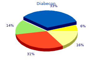
Pericarditis of renal failure occurs in up to one-third of patients with chronic uremia (uremic pericarditis) type 2 diabetes mellitus khardori cheap diabecon 60 caps buy online, is also seen in patients undergoing chronic dialysis with normal levels of blood urea and creatinine blood glucose determination order 60 caps diabecon with visa, and is termed dialysisassociated pericarditis diabetes blood glucose levels 60 caps diabecon order with mastercard. When the pericarditis of renal failure is recurrent or persistent blood sugar 2 60 caps diabecon purchase fast delivery, a pericardial window should be created or pericardiectomy may be necessary diabetes type 2 hypoglycemia buy 60 caps diabecon visa. The latter gradually contracts and forms a firm scar, encasing the heart and interfering with filling of the ventricles. In many patients the cause of the pericardial disease is undetermined, and in them an asymptomatic or forgotten bout of viral pericarditis, acute or idiopathic, may have been the inciting event. The basic physiologic abnormality in patients with chronic constrictive pericarditis is the inability of the ventricles to fill because of the limitations imposed by the rigid, thickened pericardium or the tense pericardial fluid. In constrictive pericarditis, ventricular filling is unimpeded during early diastole but it is reduced abruptly when the elastic limit of the pericardium is reached, while in cardiac tamponade, ventricular filling is impeded throughout diastole. Despite these hemodynamic changes, myocardial function may be normal or only slightly impaired in chronic constrictive pericarditis. However, the fibrotic process may extend into the myocardium and cause myocardial scarring, and atrophy, and venous congestion may then be due to the combined effects of the pericardial and myocardial lesions. In constrictive pericarditis, the right and left atrial pressure pulses display an M-shaped contour, with prominent x and y descents; the y descent, which is absent or diminished in cardiac tamponade, is the most prominent deflection in constrictive pericarditis; it reflects rapid early filling of the ventricles. They may cause few symptoms per se, and their presence may be detected by finding an enlarged cardiac silhouette on chest roentgenogram. Grossly sanguineous pericardial fluid results most commonly from a neoplasm, tuberculosis, renal failure, or slow leakage from an aortic aneurysm. Pericardiocentesis may resolve large effusions, but pericardiectomy may be required with recurrence. These hemodynamic changes, although characteristic, are not pathognomonic of constrictive pericarditis and may also be observed in cardiomyopathies characterized by restriction of ventricular filling (Chap. Supplementation of these vitamins has reduced the incidence of hyperhomocysteinemia in the United States; however, the clinical cardiovascular benefit of normalizing elevated homocysteine levels remains unproven. Hyperthyroidism Common cardiovascular manifestations of hyperthyroidism include palpitations, systolic hypertension, and fatigue. Physical examination may reveal a hyperdynamic precordium, a widened pulse pressure, increases in the intensity of the first heart sound and the pulmonic component of the second heart sound, and a third heart sound. An increased incidence of mitral valve prolapse has been described in hyperthyroid patients, in which case a midsystolic murmur may be heard at the left sternal border with or without a systolic ejection click. Elderly patients with hyperthyroidism may present with only cardiovascular manifestations of thyrotoxicosis, such as sinus tachycardia, atrial fibrillation, and hypertension, all of which may be resistant to therapy until the hyperthyroidism is controlled. Hypothyroidism Cardiac manifestations of hypothyroidism include a reduction in cardiac output, stroke volume, heart rate, blood pressure, and pulse pressure. Pericardial effusions are present in about one-third of patients, rarely progress to tamponade, and likely result from increased capillary permeability. Other clinical signs include cardiomegaly, bradycardia, weak arterial pulses, distant heart sounds, and pleural effusions. Pathologically, the heart is pale and dilated, and often demonstrates myofibrillar swelling, loss of striations, and interstitial fibrosis. In addition, obese patients have a distinct cardiovascular abnormality characterized by increased total and central blood volumes, cardiac output, and left ventricular filling pressure. The elevated cardiac output appears to be required to support the metabolic needs of the excess adipose tissue. Left ventricular filling pressure is often at the upper limits of normal at rest and rises excessively with exercise. As a result of chronic volume overload, eccentric cardiac hypertrophy with cardiac dilatation and ventricular dysfunction may develop. Pathologically, there are left and, in some cases, right ventricular hypertrophy and generalized cardiac dilatation. Weight reduction is the most effective therapy and results in reduction in blood volume and in the return of cardiac output toward normal. Treatment with angiotensin-converting enzyme inhibitors, sodium restriction, and diuretics may be useful to control heart failure symptoms. This form of heart disease should be distinguished from the Pickwickian syndrome, which may share several of the cardiovascular features of heart disease secondary to severe obesity but, in addition, frequently has components of central apnea, hypoxemia, pulmonary hypertension, and cor pulmonale. Before treatment with thyroid hormone, patients with hypothyroidism frequently do not have angina pectoris, presumably because of the low metabolic demands caused by their condition. Therefore, replacement should be done with care, starting with low doses that are increased gradually. Hypertension occurs in up to onethird of patients with acromegaly and is characterized by suppression of the renin-angiotensin-aldosterone axis and increases in total-body sodium and plasma volume. Some 50% of patients with carcinoid syndrome have cardiac involvement, usually manifesting as abnormalities of the right-sided cardiac structures. These patients invariably have hepatic metastases allowing vasoactive substances to circumvent hepatic metabolism. They occur on the cardiac valves where they cause valvular dysfunction, as well as on the endothelium of the cardiac chambers and great vessels. Carcinoid heart disease most often presents as tricuspid regurgitation, pulmonic stenosis, or both. In some cases a high cardiac output state may occur, presumably as a result of a decrease in systemic vascular resistance resulting from vasoactive substances released by the tumor. Pericardial effusions may be found echocardiographically in 1050% of patients with rheumatoid arthritis, particularly those with subcutaneous nodules. Nonetheless, only a small percentage of these patients have symptomatic pericarditis and, when present, it usually follows a benign course, only occasionally progressing to cardiac tamponade or constrictive pericarditis. Inflammation and granuloma formation may affect the cardiac valves, most often the mitral and aortic, and may cause clinically significant regurgitation owing to valve deformity. Treatment is directed at the underlying rheumatoid arthritis and may include glucocorticoids. Urgent pericardiocentesis should be performed in patients with tamponade, whereas pericardiectomy is usually required in cases of pericardial constriction. Focal myocardial necrosis and inflammatory cell infiltration are present in 50% of patients who die with pheochromocytoma and may contribute to clinically significant left ventricular failure and pulmonary edema. In addition, associated hypertension results in left 274 myocardium, resulting in aortic and mitral regurgitation, conduction abnormalities, and ventricular dysfunction. One-tenth of patients have significant aortic insufficiency and one-third have conduction disturbances; both are more common in patients with peripheral joint involvement and long-standing disease. Treatment with aortic valve replacement and permanent pacemaker placement may be required. Occasionally, aortic regurgitation precedes the onset of arthritis, and, therefore, the diagnosis of a seronegative arthritis should be considered in young males with isolated aortic regurgitation. They are most often located on the left-sided cardiac valves, particularly on the ventricular surface of the posterior mitral leaflet, and are made up almost entirely of fibrin. The lesions may embolize or become infected but rarely cause hemodynamically important valvular regurgitation. Myocarditis generally parallels the activity of the disease and, although common histologically, seldom results in clinical heart failure unless associated with hypertension. While arteritis of epicardial coronary arteries may occur, it rarely results in myocardial ischemia. Infection most commonly involves heart valves (either native or prosthetic) but may also occur on the low-pressure side of the ventricular septum at the site of a defect, on the mural endocardium where it is damaged by aberrant jets of blood or foreign bodies, or on intracardiac devices themselves. The analogous process involving arteriovenous shunts, arterioarterial shunts (patent ductus arteriosus), or a coarctation of the aorta is called infective endarteritis. Endocarditis may be classified according to the temporal evolution of disease, the site of infection, the cause of infection, or a predisposing risk factor such as injection drug use. Although each classification criterion provides therapeutic and prognostic insight, none is sufficient alone. Acute endocarditis is a hectically febrile illness that rapidly damages cardiac structures, hematogenously seeds extracardiac sites, and, if untreated, progresses to death within weeks. The risk of prosthesis infection is greatest during the first 6 months after valve replacement; gradually declines to a low, stable rate thereafter; and is similar for mechanical and bioprosthetic devices. The pathogens vary somewhat with the clinical types of endocarditis, in part because of different portals of entry. Prosthetic valve endocarditis arising within 2 months of valve surgery is generally the result of intraoperative contamination of the prosthesis or a bacteremic postoperative complication. The portals of entry and organisms causing cases beginning >12 months after surgery are similar to those in community-acquired native valve endocarditis. Transvenous pacemaker lead and/or implanted defibrillatorassociated endocarditis is usually nosocomial. The majority of episodes occur within weeks of implantation or generator change and are caused by S. Endocarditis occurring among injection drug users, especially when infection involves the tricuspid valve, is commonly caused by S. Left-sided valve infections in addicts have a more varied etiology and involve abnormal valves, often ones damaged by prior episodes of endocarditis. A number of these cases are caused by Pseudomonas aeruginosa and Candida species, and sporadic cases are due to unusual organisms such as Bacillus, Lactobacillus, and Corynebacterium species. From 515% of patients with endocarditis have negative blood cultures; in one-third to one-half of these cases, cultures are negative because of prior antibiotic exposure. Some fastidious organisms that cause endocarditis do so in characteristic epidemiologic settings. The thrombus subsequently serves as a site of bacterial attachment during transient bacteremia. These conditions result from rheumatic heart disease (particularly in the developing world, where rheumatic fever remains prevalent), mitral valve prolapse, degenerative heart disease, and congenital malformations. Fibronectin-binding proteins present on many gram-positive bacteria, clumping factor (a fibrinogen- and fibrin-binding surface protein) on S. Native valve endocarditis (whether acquired in the community or in association with health care), prosthetic valve endocarditis, and endocarditis due to injection drug use share clinical and laboratory manifestations (Table 25-2). Endocarditis caused by Staphylococcus lugdunensis (a coagulase-negative species) or by enterococci may present acutely. Extension of infection into paravalvular tissue adjacent to either the right or the noncoronary cusp of the aortic valve may interrupt the conduction system in the upper interventricular septum, leading to varying degrees of heart block. Emboli to a coronary artery may result in myocardial infarction; nevertheless, embolic transmural infarcts are rare. However, these symptoms in a febrile patient with valvular abnormalities or a behavior pattern that predisposes to endocarditis. In patients with subacute presentations, fever is typically low-grade and rarely exceeds 39. Fever may be blunted or absent in patients who are elderly or severely debilitated or who have marked cardiac or renal failure. Cardiac Manifestations Although heart murmurs are usually indicative of the predisposing cardiac pathology rather than of endocarditis, valvular damage and ruptured chordae may result in new regurgitant murmurs. Vegetations >10 mm in diameter (as measured by echocardiography) and those located on the mitral valve are more likely to embolize than are smaller or nonmitral vegetations. With effective antibiotic treatment, the frequency of embolic events decreases from 13 per 1000 patient-days during the initial week to 1. Neurologic symptoms, most often resulting from embolic strokes, occur in up to 40% of patients. Endocarditis associated with flow-directed pulmonary artery catheters is often cryptic, with symptoms masked by comorbid critical illness, and is commonly diagnosed at autopsy. Transvenous pacemaker lead and/or implanted defibrillatorassociated endocarditis may be associated with obvious or cryptic generator pocket infection and results in fever, minimal murmur, and pulmonary symptoms due to septic emboli. Late-onset prosthetic valve endocarditis presents with 279 typical clinical features. In both early-onset and more delayed presentations, paravalvular infection is common and often results in partial valve dehiscence, regurgitant murmurs, congestive heart failure, or disruption of the conduction system. Nevertheless, a highly sensitive and specific diagnostic schema- known as the Duke criteria-has been developed on the basis of clinical, laboratory, and echocardiographic findings (Table 25-3). Documentation of two major criteria, of one major and three minor criteria, or of five minor criteria allows a clinical diagnosis of definite endocarditis. The requirement for multiple positive blood cultures over time is consistent with the continuous low-density bacteremia characteristic of endocarditis (100 organisms/mL). Among patients with untreated endocarditis who ultimately have a positive blood culture, 95% of all blood cultures are positive; in 98% of these cases, one of the initial two sets of cultures yields the microorganism. Vascular phenomena: major arterial emboli, septic pulmonary infarcts, mycotic aneurysm, intracranial hemorrhage, conjunctival hemorrhages, Janeway lesions 4. Microbiologic evidence: positive blood culture but not meeting major criterion as noted previouslyb or serologic evidence of active infection with organism consistent with infective endocarditis a blood culture sets (with two bottles per set), separated from each other by at least 1 h, should be obtained from different venipuncture sites over 24 h. If the cultures remain negative after 4872 h, two or three additional blood culture sets should be obtained, and the laboratory should be consulted for advice regarding optimal culture techniques. Empirical antimicrobial therapy should not be administered initially to hemodynamically stable patients with subacute endocarditis, especially those who have received antibiotics within the preceding 2 weeks; thus, if necessary, additional blood culture sets can be obtained without the confounding effect of empirical treatment. Patients with acute endocarditis or with deteriorating hemodynamics who may require urgent surgery should be treated empirically immediately after three sets of blood cultures are obtained over several hours. Non-Blood-Culture Tests Serologic tests can be used to implicate causally some organisms that are difficult to recover by blood culture: Brucella, Bartonella, Legionella, and C. Pathogens can also be identified in surgically recovered vegetations or emboli by culture, by microscopic examination with special stains. Echocardiography Imaging with echocardiography allows anatomic confirmation of infective endocarditis, sizing of vegetations, detection of intracardiac complications, and assessment of cardiac function. It detects vegetations in >90% of patients with definite endocarditis; nevertheless, falsenegative studies are noted in 618% of endocarditis patients. Experts favor echocardiographic evaluation of all patients with a clinical diagnosis of endocarditis; however, the test should not be used to screen patients with a low probability of endocarditis. An American Heart Association approach to the use of echocardiography for evaluation of patients with suspected endocarditis is illustrated in. Blood Cultures Isolation of the causative microorganism from blood cultures is critical not only for diagnosis but also for determination of antimicrobial susceptibility and planning of treatment.
Diabecon 60 caps buy mastercard. Obstetrics - Dr.Nadine Alaa sherif - Diabetes Mellitus & Cardiac Disease in Pregnancy.
References
- Schechter MM. The superior vena cava syndrome. Am J Med Sci 1954;227(1):46-56.
- Izumi R, Shimizu K, Iyobe T, et al. Postoperative adjuvant hepatic arterial infusion of Lipiodol containing anticancer drugs in patients with hepatocellular carcinoma. Hepatology. 1994;20(2):295-301.
- Bar-Joseph G, Galvis AG: Perforation of the heart by central venous catheters in infants: Guidelines to diagnosis and management, J Pediatr Surg 18:284-287, 1983.
- Von Eiselberg A. Uber plastik bei ektropium des unterkeifers (progenie). Klin Wochenschr 1906;19:1505.
- Mustard CA, Mayer T. Case-control study of exposure to medication and the risk of injurious falls requiring hospitalization among nursing home residents. Am J Epidemiol 1997;145:738-45.