

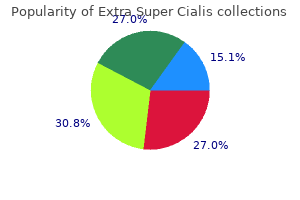
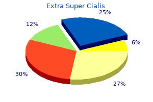
Extra Super Cialis
| Contato
Página Inicial
Thomas W. Sadler, Ph.D.
- Senior Genetics Scholar
- Greenwood Genetic Center
- Greenwood, South Carolina
In this format erectile dysfunction juice drink purchase 100 mg extra super cialis fast delivery, the cortical sulci and gyri appear in darker and lighter gray male erectile dysfunction age generic 100 mg extra super cialis mastercard, respectively erectile dysfunction causes high blood pressure purchase extra super cialis overnight delivery, on a computationally inflated surface erectile dysfunction statistics age order extra super cialis 100 mg with amex. As shown in the logo in the upper left impotence newsletter extra super cialis 100 mg buy overnight delivery, voxels that show retinotopically specific activation in the fovea are coded in red (centered at 1. Parafoveal eccentricities are shown in blue, and more peripheral eccentricities are shown in green (centered at 3. A good correlation reportedly exists between the surface and epidural recordings obtained from surgically-operated patients suffering from recurrent spreading depolarization waves due to vascular or trauma induced brain injury (Drenckhahn et al. Further research is needed to investigate these interesting possibilities in migraine patients. For example, the visual aura (experienced as a contralateral visual hallucination) and hemi-cranial pain (experienced most often ipsilateral to the dysfunctional hemisphere, and a source for ipsilateral trigeminovascular activation) suggests that the occipital perturbation underlying the aura could also cause headache (Olesen et al. Degranulated mast cells (f) can easily be distinguished from resting cells (e) with loss of blue cytoplasmic granules. The mouse and rat grimace scales demonstrated that rodents surprisingly, have facial expressions displaying their discomfort during pain (Langford et al. However, such activation is expected to lead to a transient headache during the aura, not 1520 minutes later. This brief elevation may contribute to the headache seen simultaneously with the beginning of aura (Russell and Olesen, 1996; Hansen et al. Unfortunately, clinical studies investigating the relationship between the aura and headache onset did not consider changes in the intensity and characteristics of the headache, which are known to vary over time (see Chapter 11). The signaling cascade began 16 From cortical spreading depression to trigeminovascular activation in migraine 273 Mid dle M ge enin al A rter y ate am Dur tery l Ar Pia r. Reproduced with the permission of the American Association for the Advancement of Science. However, there are still controversies about the generalizability of each of these factors detected under specific in vitro conditions to neurons in situ. Under resting conditions, the opening probability of pannexin-1 channels is low (or opens in a low conductance state), possibly because their unusually large conductance, if opened, may jeopardize the physiological transmembrane ion gradients. The increase was maximal at the first-hour blood sampling point, and then declined. Since the internal jugular vein in humans drains mainly the brain parenchyma (unlike the internal jugular vein in the rat, which is a rather thin vessel compared to the external jugular vein, which mainly provides the cerebral venous drainage (Szabo, 1995)), these observations suggest production of cytokines in brain parenchyma during a migraine attack as the above outlined hypothesis proposed (Karatas et al. By contrast, cytokine measurements in peripheral blood during migraine attacks, despite several positive reports, have generally led to inconclusive results, possibly due to plasma volume dilution of relatively small absolute increases in cytokine levels, even in jugular blood, plus the timing of blood sampling (Durham and Papapetropoulos, 2013). These patients also experience rash, fever, malaise, arthralgia, myalgia, conjunctivitis and non-migrainous headache usually induced by cold. However, caution is needed in interpreting these observations in favor of activation of the parenchymal pathway. Further supporting this view, headaches directly starting from meninges, such as during subarachnoid hemorrhage or meningitis, show several characteristic features of migraine headache, including its throbbing nature and photo/phonophobia, and sometimes they are alleviated by sumatriptan (Lamonte et al. Experimental data have not only established the presence of large networks of neuropeptide-containing meningeal afferents in most vertebrates, including man (See Chapter 1), but calcium-dependent neuropeptide release from these meningeal fibers (Moskowitz et al. Although, to date, these meningeal networks have received most attention for their role in migraine, in other tissues, neurogenic inflammation is part of an integrative protective response between the peripheral nervous system and the adaptive and innate immune systems, in response to tissue danger signals (Chiu et al. The parenchymal and meningeal inflammatory signaling cascades seem to be tightly regulated, as they do not lead to overt inflammation characterized by infiltration of leukocytes. The rapid decline in the elevated level of inflammatory mediators in the internal jugular vein of migraine patients supports this idea (Sarchielli et al. It is likely that the algesic signals arise from the parenchymal inflammatory cascade, and are amplified by the meningeal neurogenic inflammation, which may sustain the activation of the nociceptors for development of the sensitization and a lasting headache (see Chapters 6 and 7). The region of increased uptake three hours after the injection corresponded to the diagram where the subject localized her migraine pain epicenter. This conforms with the idea that there may be a threshold to translate the parenchymal stress to neurogenic inflammation and lasting headache. Furthermore, there is a small group of patients who experience only aura without headache (Headache Classification Committee of the International Headache Society, 2013). A reduced volume of depolarized tissue may, therefore, decrease its algesic potential by reducing the concentrations of cytokines reaching the subarachnoid space, although they may still cause a spreading sensory percept (see also Chapter 17). Moreover, some people may have a higher headache threshold for a number of reasons, as the presence of people who never suffer from headache suggests. For example, their parenchymal inflammatory response may not be strong enough to initiate the meningeal neurogenic inflammation, or the neurogenic inflammation may have a higher threshold. The neuronal stress sensed by pannexin-1 large-pore channels, and conveyed by the inflammatory signaling to be reported as headache, may also be experienced during intense neuronal activity under suboptimal homeostatic conditions. Prevalence of typical migraine aura without headache in Japanese ophthalmology clinics. Does single cortical spreading depression elicit pain behaviour in freely moving rats Sensitization of the trigeminovascular pathway: perspective and implications to migraine pathophysiology. Indomethacin and acetylsalicylic acid block neurogenic plasma protein extravasation in rat dura mater. Long-term elevation of cyclooxygenase-2, but not lipoxygenase, in regions synaptically distant from spreading depression. Cognitive disturbances and regional cerebral blood flow abnormalities in migraine patients: their relationship with the clinical manifestations of the illness. A voxel-based analysis of brain activity in high-order trigeminal pathway in the rat induced by cortical spreading depression. Migraine generator network and spreading depression dynamics as neuromodulation targets in episodic migraine. Cortical hot spots and labyrinths: why cortical neuromodulation for episodic migraine with aura should be personalized. Correlates of 280 Neurobiological Basis of Migraine spreading depolarization in human scalp electroencephalography. Biomarkers associated with migraine and their potential role in migraine management. Cortical Spreading Depression Causes Unique Dysregulation of Inflammatory Pathways in a Transgenic Mouse Model of Migraine. Immunomodulatory Effect of Toll-Like Receptor-3 Ligand Poly I: C on Cortical Spreading Depression. Proceedings of the National Academy of Sciences of the United States of America 98(8): 468792. The role of nitric oxide in the development of cortical spreading depression-induced tolerance to transient focal cerebral ischemia in rats. Optogenetic induction of cortical spreading depression in anesthetized and freely behaving mice. Cortical spreading depression induces proinflammatory cytokine gene expression in the rat brain. Supraphysiological whisker stimulation initiates an inflammatory response in the mouse barrel cortex in vivo. Hippocampal spreading depression bilaterally activates the caudal trigeminal nucleus in rodents. Clinical relevance of cortical spreading depression in neurological disorders: migraine, 282 Neurobiological Basis of Migraine malignant stroke, subarachnoid and intracranial hemorrhage, and traumatic brain injury. Pharmacological characterization of pannexin-1 currents expressed in mammalian cells. Note on a possible correspondence between the scotomas of migraine and spreading depression of Leao. Critical role for mast cells in interleukin-1beta-driven skin inflammation associated with an activating mutation in the nlrp3 protein. Timing and topography of cerebral blood flow, aura, and headache during migraine attacks. Pearls and pitfalls in experimental in vivo models of headache: conscious behavioral research. Ergot alkaloids block neurogenic extravasation in dura mater: proposed action in vascular headaches. Chemokine levels in the jugular venous blood of migraine without aura patients during attacks. Proinflammatory cytokines, adhesion molecules, and lymphocyte 284 Neurobiological Basis of Migraine integrin expression in the internal jugular blood of migraine patients without aura assessed ictally. Mechanisms of spreading depression and hypoxic spreading depression-like depolarization. The Rat Grimace Scale: a partially automated method for quantifying pain in the laboratory rat via facial expressions. The cranial venous system in the rat: anatomical pattern and ontogenetic development. Temporal profiles of high-mobility group box 1 expression levels after cortical spreading depression in mice. Minimum conditions for the induction of cortical spreading depression in brain slices. Brief report: bilateral spreading cerebral hypoperfusion during spontaneous migraine headache. Cyclooxygenase-2 expression associated with spreading depression in a primate model. In general, computational neuroscience uses abstract models formulated in mathematical language to interpret data and guide a principled understanding of the nervous systems in health and disease. The complexity of neural systems can make it difficult to assess the value of such models, because they have to abstract from the details and incorporate only the aspects that are considered to be important, yet what is important may only be known in hindsight. With regard to this difficulty, however, a computational model is often not fundamentally different from animal models. In particular, in animal models of pain, the efficacy of a drug is often first demonstrated in humans and then in the animal model, which is called backward validation [41]. Unlike animal models, a computational model can be deliberately and intentionally changed in any manner that is, components can be modified or added in any sequence. Therefore, one can feature backward validation by making the computational science process cyclic. Moreover, experiments on pain using non-human animals, or even human subjects pose great ethical problems. Last, but not least, experimental animal models and clinical studies have become increasingly sophisticated, and have given rise to an abundance of biological data across a range of spatial and temporal scales, extending from molecules to whole brain, and from sub-milliseconds of channel gating to several hours of attack duration, and even years in changes in headache history. Accurate biophysical, multi-scale computational models can help to integrate diverse data sets and to unify information. The common theme that runs through these examples is to elucidate how the respective objective of the model determines the chosen level of abstraction. First, we introduce microscopic models that include cellular and cytoarchitectonic detail. Such models can address questions related to the electrophysiological Neurobiological Basis of Migraine, First Edition. In this case, the computational science process begins by identifying the following physiological observations to be selected for a working model. The threshold can initially be defined as the minimal current amplitude of, in principle, infinite duration (in practice, several tens of milliseconds suffice), that results in periodic firing the so-called rheobase current. When positive electric charge (such as sodium ions) flows into neurons, this defines an inward current. The step towards a working model is the simplification of the observed cellular system into its quantifiable factors. Among these quantifiable factors are five active membrane currents, two passive electrical properties (leak currents), and active pumping of sodium ions (Na+) and potassium ions (K+). This framework formulates membrane electrophysiology, considering the equivalent electrical circuit for a patch of membrane as a fixed capacitance (lipid bilayer) and, in parallel, voltage-gated conductances (ion channels) with, in series, batteries (ion gradients). The voltage-gated ion channel kinetics are described by independent conformational transitions of the channel subunits. It is worthwhile to look into this more fully, due to the great significance and relationship between action potentials and spreading depression. First, cellular transmission of action potentials is one of the most fundamental processes in biology. Second, far beyond the generation of action potentials is the phenomenon of excitability. How this is related to the onset of rhythmic activity, such as bursting, is important to a whole host of other problems outside membrane physiology and even biology, including physics, chemistry, sociology, and engineering. For this reason, we explicitly note that action potentials and spreading depression share many elements that constitute two distinct and separate kinds of excitable phenomena. Excitability means that, while small perturbations return immediately to rest, a stronger but still brief stimulus can trigger a large excursion that, eventually, also reverts to the resting state without the need of further input. An action potential is caused in the neural membrane by a small amplitude depolarization. By definition, the size of the rheobase current can be taken as an inversely proportionate measure of this kind of membrane excitability. In the case of an action potential, the "excursion", as an essential act constituting excitability, is the change of the membrane potential towards a positively polarized state that eventually repolarizes to the negatively polarized resting state. During this period, ions are exchanged across the nerve cell membrane, but only to a negligible degree. Spreading depression, on the other hand, is caused in the neural membrane by sustained depolarization. High-frequency discharges caused by an applied depolarizing stimulation current of 0. The orange and yellow bar at the top mark the distinct phases in the "plane of excitability". The plane is spanned by extracellular potassium ion concentration [K+]e and potassium ion clearance (measured in mM with virtual reference to the extracellular volume).
Ardraka (Ginger). Extra Super Cialis.
- Is Ginger effective?
- Nausea and vomiting following surgery.
- Preventing motion sickness and seasickness.
- What other names is Ginger known by?
- How does Ginger work?
Source: http://www.rxlist.com/script/main/art.asp?articlekey=96924
Other contributors to a lesser degree were psychologic stress erectile dysfunction doctor in delhi discount extra super cialis online amex, anxiety erectile dysfunction vacuum discount 100 mg extra super cialis overnight delivery, obsessive-compulsive feelings erectile dysfunction doctors raleigh nc cheap extra super cialis 100 mg on line, and pain coping strategies erectile dysfunction doctor boca raton buy cheap extra super cialis 100 mg. Arthritis this is pain of joint origin with clinical characteristics of inflammation or infection: edema erectile dysfunction treatment needles purchase genuine extra super cialis line, erythema, and/or increased temperature. Associated symptoms can include occlusal changes such as ipsilateral posterior open bite if intra-articular swelling is present unilaterally. This disorder has also been referred to as synovitis or capsulitis; however, these terms limit the sites of nociception. With this localized condition, there should be no history of systemic inflammatory disease. Joint pain is characterized by localized pain that is exacerbated by function and parafunction. On occasion, there may be a fluctuating swelling that decreases the ability to occlude on the ipsilateral posterior teeth. The patient is negative for rheumatologic disease, including those in systemic arthritides. Although not required for this diagnosis and without diagnostic validity, elimination of the opening and closing noise, if present, with protrusion can help corroborate this diagnosis. It is characterized by an abnormal relation or misalignment of the articular disc relative to the condyle. Although posterior and mediolateral displacements of the articular disc have been described, the usual direction for displacement is in an anterior or anteromedial direction. When the disc does not reduce with opening of the mouth, intermittent limited mandibular opening occurs. Although not required for this diagnosis, occurrence of intermittent closed lock during the clinical examination can help corroborate this diagnosis. History is positive for both of the following: · In the last 30 days or during the examination itself, any noises present with jaw movement or function · In the last 30 days, report of intermittent locking with limited opening or evidence of intermittent locking during clinical examination Examination is positive for the following: · Disc displacement with reduction as defined above When this diagnosis needs to be confirmed, the imaging analysis criteria are the same as for disc displacement with reduction. If locking occurs during imaging, then an imaging-based diagnosis of disc displacement without reduction will be rendered, and clinical confirmation of reversion to intermittent locking is needed. This disorder is associated with limited mandibular opening that does not reduce with the clinician or patient performing a manipulative maneuver. History is the same as defined for disc displacement without reduction with limited opening. Examination is positive for the following: · Maximum assisted opening (passive stretch) > 40 mm including vertical incisal overlap When this diagnosis needs to be confirmed, then imaging analysis criteria are the same as for disc displacement without reduction with limited opening. In case of bilateral involvement, asymmetries in mandibular movements during clinical examination will be less pronounced or absent. The diagnostic criteria of mandibular movement asymmetries are for unilateral causes of hypomobility. Hypomobility is firm and unyielding because of intra-articular fibrous adhesions, more widespread fibrotic changes in the capsular ligaments (fibrous ankylosis), and/or, less 158 frequently, the formation of a bony mass that results in fusion of the joint components (bony ankylosis). Adhesions may occur secondary to joint inflammation that results from direct trauma, excessive loading, or systemic conditions such as a polyarthritic disease. It is characterized by radiographic evidence of bone proliferation with marked deflection to the affected side and marked limited laterotrusion to the contralateral side. Note that the condyle is frequently anterior to the eminence at full mouth opening and thus by itself is not a predictor of hypermobility disorders. Pain may occur at the time of dislocation with residual pain following the episode. In the open mouth position, the disc-condyle complex is positioned anterior to the articular eminence and is unable to return to normal closed mouth position without a manipulative maneuver by the patient. When the patient needs the assistance of the clinician to reduce the dislocation and normalize the jaw position, it is called luxation or open lock. If the patient presents with this disorder, he or she is then found clinically positive for inability to return to a normal closed mouth position without manipulative patient maneuver. The condition occurs spontaneously, is mainly bilateral, and occurs primarily in adolescent and young adult females. In early stages, dental occlusal changes may not be evident, but imaging findings would be positive. Serologic tests for rheumatologic disease, including systemic arthritides, must be negative. In addition, laboratory testing will confirm negative serologic results for rheumatologic disease. They can vary from no signs or symptoms to only pain to only swelling and exudate to only tissue degradation to only growth disturbance. Resorption of condylar structures may be associated with malocclusion such as a progressive anterior open bite. A diagnostic instrument should aim to identify patients with chronic inflammation early and accurately, should not exclude patients with chronic arthritis of long duration, and should not only cover rheumatoid arthritis but the whole range of chronic inflammatory states. Its main characteristic is the formation of cartilaginous nodules that may be pedunculated and/or detached from the synovial membrane, becoming loose bodies within the joint space. The disease may be associated with malocclusion, such as a progressive ipsilateral posterior open bite. Fractures Direct traumatic force can injure all related bony components of the masticatory system (ie, temporal bone, maxilla, zygoma, sphenoid bone, and mandible). This trauma can be related to the following conditions: fracture, dislocation, contusion, or laceration of articular surfaces, ligaments, and disc, with or without intraarticular hemarthrosis. Sequelae could include adhesions, ankylosis, occlusal abnormalities, or joint degeneration. History must be positive for both of the following: · Progressive development of mandibular asymmetry or micrognathia from birth or early childhood · Development of malocclusion, which may include posterior open bite Examination is positive for both of the following: · Confirmation of mandibular asymmetry, with deviation of the chin to the affected side or micrognathia · Inability to detect the condyle upon palpation during mandibular movements. It is commonly associated with other congenital anomalies (eg, oculo-auriculo-vertebral spectrum [Goldenhar syndrome], hemifacial microsomia, and mandibulofacial dysostosis [Treacher Collins syndrome]). It is occasionally bilateral, and in such cases asymmetry is not present, but micrognathia is the dominant clinical manifestation. It can occur unilaterally or bilaterally as a localized enlargement such as condylar hyperplasia, or as an overdevelopment of the entire mandible or side of the face. To diagnose hyperplasia, the history must be positive for progressive development of mandibular or facial asymmetry, and the examination must confirm this history. Fibromyalgia is of particular interest because it may easily be confused with a regional masticatory muscle disorder. Although not required in the criteria, a positive finding with the specified provocation tests when examining the other masticatory muscles can help to corroborate this diagnosis if the location of the pain associated with the chief complaint is in these muscles; limited mandibular range of motion may also be present. History must be positive for both of the following: · Pain in the jaw, temple, ear, or in front of the ear in the last 30 days. Masticatory muscle disorders the mechanisms that produce pain in skeletal muscles are still not well understood. Overuse of a normally perfused muscle or ischemia of a normally working muscle may cause pain. Endogenous substances (eg, bradykinin, serotonin, prostaglandins, neuropeptides, and substance P) can sensitize the nociceptive endings very easily. Local myalgia this involves pain of muscle origin plus a report of pain localized only to the site of palpation (immediate site to stimulation). The criterion for this diagnostic group limits the familiar pain to be provoked only with palpation and not with mandibular movement. Myofascial pain with spreading this involves pain of muscle origin plus a report of pain spreading beyond the location of the palpating fingers but within the boundary of the masticatory muscle being examined. To diagnose myofascial pain with spreading, the patient must have local myalgia, and the examination of the temporalis or masseter muscle must confirm both of the following: · Familiar muscle pain with palpation · Pain with muscle palpation with spreading of the pain beyond the location of palpation but within the boundary of the palpated muscle Note: Other masticatory muscles may be examined as required. Myofascial pain with referral this involves pain of muscle origin as defined for myalgia plus a referral of pain beyond the boundary of the masticatory muscles being 166 palpated, for example, to the ear, tooth, or eye; spreading may be present. To diagnose myofascial pain with referral, the patient must have myalgia, and the examination of the temporalis or masseter muscle must confirm both of the following: · Familiar muscle pain with palpation · Pain with muscle palpation beyond the boundary of the muscle Other masticatory muscles may be examined as required. The temporalis tendon is a common site of tendonitis with referred pain to the teeth or other structures. To diagnose tendonitis, the patient must have myalgia as previously defined, and the examination must confirm the diagnosis of myalgia but restricted to the temporalis tendon. It generally arises acutely following direct trauma of the muscle or from infection or chronically with autoimmune disease. To diagnose a spasm, the patient must report the following: · Immediate onset of muscle pain modified by function and parafunction as operationalized in myalgia · Immediate report of limited range of jaw motion In addition, the examination must confirm both of the following: · Myalgia that may include all masticatory muscles · Limited range of jaw motion in the direction that elongates the affected muscle (eg, for jaw closers, opening will be limited to < 40 mm; for the lateral pterygoid muscle, ipsilateral movement will be limited to < 7 mm) When this diagnosis needs to be confirmed, laboratory testing will confirm elevated electromyographic activity compared with the contralateral unaffected muscle. To diagnose contracture, the patient must have progressive loss of range of motion, and the examination must confirm that unassisted and assisted jaw movements are limited such that maximum assisted opening is < 40 mm. It must also be associated with a hard end feel that has been operationalized as having unyielding resistance to assisted movements as felt by the clinician. It is usually not associated with pain and can be secondary to overuse and/or chronic tensing of the muscle(s). To diagnose hypertrophy, the patient must have enlargement of one or more masticatory muscles as evidenced from photographs or previous records, and the examination must confirm this enlargement. They may present with swelling, spasm, pain during function, limited mouth opening, and/or sensory-motor changes (eg, paresthesias). They could be components of a number of central nervous systems disorders, including Parkinson disease and Meige syndrome, and could be an adverse event related to medication usage, notably neuroleptics. Trauma to the brain, head, and neck can trigger the onset of transient or permanent dystonia of the masticatory muscles. The condition can make opening and closing the mouth difficult and impair speech, swallowing, and chewing. To allocate these diagnoses, the patient must provide a positive history of neurologic diagnosis of dystonia in the orofacial region and history components of myalgia and/or arthralgia (diagnostic criteria as previously described) that worsens with episodes of dystonia. It is more common with advancing age and in patients with a history of using neuroleptic medications and/or associated with traumatic brain injury, psychiatric conditions, or other neurologic disorders (eg, Wilson disease). Reduction or discontinuation of the movement pattern could occur when the mouth or face receives sensory stimulation (sensory trick). To allocate these diagnoses, the patient must provide a positive history of neurologic diagnosis of dyskinesia in the orofacial region and history components of myalgia and/ or arthralgia (diagnostic criteria as previously described) that worsens with episodes of dyskinesia. History and examination are positive for both a rheumatologic-based diagnosis of fibromyalgia and myalgia as previously defined. Historically, in diagnosing fibromyalgia, there needed to be tenderness in at least 11 of 18 specified sites and the presence of widespread pain denoted as axial pain, right and left pain, and upper and lower segment pain. Current criteria eliminate the tender points and focus on a widespread pain index and symptom severity scale. Intermittent muscle pain conditions may not produce centrally mediated myalgia, while a prolonged and constant period of muscle pain is likely to lead to the condition. They include decreased pain, decreased adverse loading, restoration of function, and resumption of normal daily activities. These management goals are best achieved by a well-defined program designed to treat the physical disorder(s) and to reduce or eliminate the effects of all contributing factors. Therefore, special efforts should be made to avoid early use of aggressive, irreversible treatments such as complex occlusal therapy or surgery. To diagnose coronoid hyperplasia, the patient must complain of progressive limitation of jaw opening, the examination must confirm the reduction of active and passive maximum jaw opening, and imaging must show an elongated coronoid process that approximates the posterior aspect of the zygomatic process of the maxilla on opening. Therefore, in addition to the physical diagnosis, the goal of each evaluation should be the development of a prioritized problem list of the relevant contributing factors. Early treatment of acute musculoskeletal pain results in greater patient satisfaction, fewer work days lost, and reduced chance of development of a chronic pain condition. Reasons for this vary, but these patients typically fall into two groups: patients with an incomplete or incorrect diagnosis or patients with unsuccessfully addressed or even unrecognized contributing factors. It is difficult for an individual clinician to address the multiple contributing factors that may be present in complex chronic pain patients. Patient education and self-management the success of a self-management program depends on patient acceptance, motivation, cooperation, and compliance. The time spent on patient reassurance and education is a significant factor in developing a high level of rapport and treatment compliance. A successful self-management program allows healing and prevents further injury to the musculoskeletal system. Modification of function (eg, avoidance of heavy mastication, gum chewing, wide yawning, singing, and playing certain musical instruments) and parafunctional habit reversal (eg, clenching, bruxing, tongue thrusting, cheek biting, poor sleeping posture, or object biting) should be emphasized. Offending habits can be modified with habit awareness, motivation to change, and knowledge in how to change. Commitment to conscientious monitoring for the habits can lead to successful habit modification. Keeping a diary aimed at identifying circumstances and activities that foster the offending habits may also be helpful so that the patient can titrate the need for more or less monitoring during any given day. Progress with habit modification should be discussed at each follow-up appointment with the patient. A program of heat and/or ice to the affected areas, massage of the affected muscles, and gentle range-ofmotion exercises can decrease tenderness and pain and increase range of motion. Heat should not be used following an acute injury (ie, less than 72 hours) or for acute inflammation or infection. Ice packs are used primarily for local analgesic and anti-inflammatory ef172 fects in muscle and joint tissues. Because the temperature differential is greater with cold, a shorter application may produce a greater response. Cold should not be used over areas with poor circulation (eg, as a result of diabetes or radiation) or over open wounds. Recently, efforts were taken to construct an expert-based standardized definition of self-management. The core components identified for self-management include education; self-exercises; self-massage; thermal therapy; dietary advice and nutrition; and parafunctional behavior identification, monitoring, and avoidance.
C Heart failure Vein of Galen malformations present in neonates with evidence of high output heart failure erectile dysfunction non prescription drugs purchase discount extra super cialis on line. D Carotid-cavernous fistula Traumatic carotid-cavernous fistulae can occur after motor vehicle accidents or other intracranial trauma erectile dysfunction treatment in egypt purchase extra super cialis overnight delivery. They present with orbital pain erectile dysfunction age 36 buy extra super cialis 100 mg cheap, chemosis erectile dysfunction treatment pakistan cheap extra super cialis 100 mg without prescription, proptosis erectile dysfunction pills gnc discount extra super cialis 100 mg otc, ophthalmoplegia, and visual loss. Patients should undergo vascular imaging and may require interventional or surgical treatment of the fistula. B False Radiation therapy damages cells by firing particles into an atom and releasing free electrons causing damage downstream. In fully oxygenated cells, oxygen combines with unpaired free electrons to form peroxides, which are more stable and lethal than free radicals, and therefore an oxygenated cell is more sensitive to damage by radiation therapy. B > 3 years Children less than 3 years of age are particularly sensitive to cranial radiation and can have severe developmental side effects. Children should be greater than 3 years of age to be eligible for cranial radiation. B 3 cm or less Gamma knife radiosurgery can be useful for cranial masses, but should be reserved for patients with brain tumors that are 3 cm or less in maximum diameter. This size cutoff decreases the risk of harmful radiation side effects to surrounding brain structures. B 10 Gy Safe doses of radiation to the optic apparatus are generally thought to be 8 to 10 Gy. C 10 Gy or less the lens of the eye can tolerate 10 Gy or less radiation with minimal side effects. D 40 to 50% At 10 years posttreatment, approximately 40 to 50% of patients who receive sellar radiation will experience hypopituitarism as a side effect from radiation. B 4 to 6 Gy Based on current literature, the mean safe radiation dose to the cochlea is considered to be approximately 4. There is some controversy on this topic currently, but based on data available, doses from 4 to 6 Gy should be considered optimal. C Whole brain radiation Current literature supports the use of whole brain radiation in patients who have undergone resection of a cerebral metastasis. Doses up to 50 Gy have been shown to control > 90% of micrometasases, but at this dose there is a very high chance of early radiation side effects. B 10 or less Based on current studies, up to 10 concurrent cerebral metastases can be treated with stereotactic radiosurgery with good outcome and low risk of side effects. There are surgeons who feel that even this number can be safely extended, research is pending. Radiation works by causing damage to the endothelium and ultimately causing fibrosis. This process can take 2 to 3 years to develop, so risk of hemorrhage needs to be discussed with the patient over that treatment timeframe. Higher radiation doses have been associated with an increased risk of complications and no significant improvement in obliteration rates. D Dementia Dementia is the main complication from whole brain radiation after use for intracranial metastases. Incidence has been shown to be higher when patients receiving doses of 25 to 39 Gy receive those doses in fractionations that are > 300c Gy Further Reading: Greenberg. A 8 Gy Emergency radiation can be delivered to radiosensitive spine tumors when there is evidence of compression. In many circumstances, an initial dose of 8 Gy will be given to shrink the tumor, followed by further fractionated radiation after the acute situation has resolved. C 30 Gy in 10 fractions Radiation to the spine for metastatic disease in the setting of radiosensitive tumors is often administered at a dose of 30 Gy delivered over 10 fractions. A Anesthesia dolorosa Anesthesia dolorosa is a feared complication of intentional damage to the trigeminal nerve. It occurs after damage to the V1 segment of the nerve, and can lead to anesthesia of the cornea, causing patients to get recurrent corneal abrasions. Initially, imaging 186 8 Neurosurgery of the brain should be performed to rule out mass lesions or evidence of multiple sclerosis. A Start carbamazepine this patient appears to have symptoms consistent with trigeminal neuralgia. Initially, imaging of the brain should be performed to rule out mass lesions or evidence of multiple sclerosis. It is an excellent option for patients who can tolerate a small craniotomy and have a life expectancy of longer than 5 years. It is important to determine the difference between the two as treatment is different. B Fludrocortisone Fludrocortisone acts directly on renal tubules to increase sodium absorption and can be a useful medication adjunct when treating cerebral salt wasting. A Normal saline infusion In cerebral salt wasting, patients are hypovolemic and hyponatremic. Fluid resuscitation with normal saline at 100 to 125 mL/hr should be instituted in an attempt to normalize fluid status. B Severe dehydration the main complication of untreated diabetes insipidus is severe dehydration Further Reading: Greenberg. A Drink to thirst In an awake, conscious and ambulatory patient with mild diabetes insipidus, sodium levels should be monitored, but patients should be allowed to drink to thirst. D 72 hours Dobutamine increases cardiac output by positive inotropy, but patients will exhibit tachyphylaxis after approximately 72 hours of administration. B 5 to 10K One unit of platelets (out of the standard six pack) will raise the platelet count approximately 5 to 10K. A 10K In the absence of evidence of bleeding, platelets should be transfused prophylactically when the count drops to 10K. B 1 mg protamine/100 u heparin Protamine sulfate can be used to reverse the effects of unfractionated heparin, and should be administered in doses of 1 mg protamine/100 u heparin. B 4 hours Idarucizumab (Praxbind) is an effective reversal agent for the direct thrombin inhibitor Dabigatran (Pradaxa). C Malignant hyperthermia Malignant hyperthermia can occur after anesthetic use of volatile anesthetics, specifically halothane, or muscle relaxants such as succinylcholine. C Dantrolene Malignant hyperthermia can occur after anesthetic use of volatile anesthetics, specifically halothane, or muscle relaxants such as succinylcholine. B Ryanodine Malignant hyperthermia can occur after anesthetic use of volatile anesthetics, specifically halothane, or muscle relaxants such as succinylcholine. It is thought to occur in some cases due to genetic defects in the ryanodine receptor on the sarcoplasmic reticulum. B 3% or less Current literature suggests that you should have a 3% or less overall complication rate to justify performing a carotid endarterectomy. C Blood pressure control this patient is likely experiencing cerebral hyperperfusion syndrome given that blood flow to the ipsilateral hemisphere has now greatly increased. This is a controversial area, but close blood pressure control can help decrease the symptoms of cerebral hyperperfusion syndrome. A Hypoglossal palsy the distal hypoglossal nerve is often seen during the dissection for a carotid endarterectomy and a postoperative palsy has been reported to be as high as 8% in some series. Care should be taken to avoid damaging the hypoglossal nerve during the dissection. B Bedside decompression this patient has an obvious arteriotomy closure disruption and it is causing tracheal deviation and respiratory compromise. In many practices, surgeons utilize carotid angioplasty and stenting in patients with high-riding carotid bifurcations or very difficult appearing stenosis that might have a higher rate of operative complications. B Anterior and posterior cortex A spinal fracture is considered a burst fracture when both the anterior and posterior cortices of the vertebral body are violated. B Burst fracture A spinal fracture is considered a burst fracture when both the anterior and posterior cortices of the vertebral body are violated. This treatment technique is based on the Kirkham Wood study evaluating asymptomatic burst fractures managed with bracing alone. Some surgeons are avoiding bracing completely in these patients, and this is a somewhat controversial issue that is still undergoing further research. A Anterior cortex only A compression fracture of the spine occurs when imaging demonstrates violation of the anterior cortex only. A Compression fracture A spinal fracture is considered a compression fracture when the anterior cortex is violated but other aspects of the spine remain intact. A Flexion compression this image demonstrates a compression fracture, evidenced by violation of only the anterior cortex. This injury occurs via a flexion compression loading mechanism with the axis of rotation located within the anterior vertebral body. B Pure axial load this image demonstrates a burst fracture (violation of both the anterior and posterior cortices). C Flexion distraction this image demonstrates a three column pure bony injury, often referred to as a Chance fracture after the physician G. It is caused by a flexion distraction force mechanism where the axis of rotation is anterior to the vertebral body. D Shear this image demonstrates a severe spinal cord fracture caused by shear injury to the spinal column. The forces applied to the spine in order to achieve this level of dislocation are significant. C Extension distraction this image demonstrates a three column spine fracture in a patient with evidence of ankylosis of the spine. While these patients form bone easily, 190 8 Neurosurgery the bone within the vertebral body is brittle and fractures easily. This fracture was caused by an extension distraction mechanism with the axis of rotation within the posterior spinal elements. C T12-L1 Burst fractures are caused by pure axial load mechanisms and most often occur in the T12-L1 region since the T12-L1 vertebral bodies are the most centered in the midline between the lordotic lumbar spine and kyphotic thoracic spine. It is in this region where the force vector in a pure axial load mechanism is applied directly through the center of the vertebral body causing a dispersion fracture mechanism leading to disruption of the anterior and posterior vertebral cortices causing a burst fracture to occur. In this setting, the patient gets 2 points for burst fracture, 2 points for indeterminate ligamentous status and 2 points for nerve root injury, leading to a score in the operative treatment range. B Middle column In the 3-column model of Denis, the posterior vertebral body is located within the middle column. C Posterior column In the 3-column model of Denis, the facet joints are located within the posterior column. A Stable Isolated transverse process fractures of the thoracolumbar spine are generally considered stable when not associated with other major injuries and can be managed expectantly. Specific rates depend on fracture characteristics including displacement and angulation, but 30% is a good rule to consider when deciding management. More than 3 mm and the integrity of the transverse ligament should be evaluated to determine stability. It is measured by the length of a line drawn from the basion to the posterior arch of C1 divided by a line drawn from the opisthion to the anterior arch of C1. D Atypical this axis fracture is one of the atypical variety given that the fracture occurs through the base of the C2 vertebral body and the posterior cortex of the body is left in place rather than fracturing through the pedicles of C2. This fracture type should be evaluated closely as the spike of remaining vertebral body can cause the spinal cord to be 192 8 Neurosurgery draped over the shard anteriorly and cause spinal cord injury. They should be recognized as they are a subset of fractures that should not be put in traction as this can worsen the injury type. There could be an underlying spinal cord injury and reduction with closed traction should be performed. The superior articulating process on the left is dorsal to the inferior articulating process and there is evidence of rotation. If any change in the neuro exam occurs, the weight can be removed and the exam re-confirmed. Atlas of Neurosurgical Techniques: Spine and Peripheral Nerves, 2nd edition, 2016, page 237. If the patient is awake, an exam should be performed as well after the addition of any weight. C Transverse ligament the rule of Spence is calculated be measuring the displacement of the C1 lateral masses on the C2 joints. B 7 mm the rule of Spence is calculated be measuring the displacement of the C1 lateral masses on the C2 joints. If the total overhang of the C1 lateral masses on the C2 joints is > 7 mm, disruption of the transverse ligament should be suspected. Scar tissue will enhance homogeneously while a recurrent disk may peripherally enhance but will not demonstrate homogeneous enhancement. C 85% Approximately 85% of patients with a herniated lumbar disc will improve with 6 weeks of nonoperative management, thus an initial period of non-surgical management should be attempted before surgical decompression is considered. This does not include patients with progressive weakness or cauda equina syndrome. When full saddle anesthesia 194 8 Neurosurgery develops, few patients will return to normal bladder function Further Reading: Greenberg. C ~ 75% Approximately 73% of patients will be free of leg pain 1 year after surgery for a herniated lumbar disc. B 4% at 10 years According to current literature, the rate of recurrent disc herniation at the same level on either side is approximately 4% at 10 years, with 1/3rd of those patients experiencing the recurrence within the 1st year postoperation. To evaluate for a pseudoarthrosis, measurements should be taken between the spinous processes at that level.
Because of the serious implications of the diagnoses and the nonspecific nature of the common presenting complaints best erectile dysfunction pills treatment extra super cialis 100 mg with mastercard, a disciplined erectile dysfunction causes diabetes order extra super cialis 100 mg on-line, systematic approach to the consideration of these disorders is necessary erectile dysfunction doctor in karachi purchase generic extra super cialis on line. The list of disorders included in the inherited arrhythmia syndromes varies impotence back pain discount extra super cialis 100 mg buy, and it has evolved over the last 20 to 30 years cough syrup causes erectile dysfunction buy generic extra super cialis 100 mg line, reflecting the ongoing recognition, definition, and characterization of these diseases. In this discussion, we will include six disorders in two general groups: ion channelopathies and diseases of the myocardium. Owing to their rarity and/or their association with other, more prominent, noncardiac abnormalities, these disorders are not discussed in detail here. For an electrophysiologist who sees patients with these syndromes and obtains genetic testing, an understanding of basic genetics is essential to accurately interpret results and provide accurate advice and counseling. In this section, we will discuss the basics of genetics as they apply to these syndromes and review the relevant nomenclature and available genetic tests. The human genome is composed of over 4 billion bases that make up approximately 21,000 genes arrayed on 23 chromosomes. Human cells are diploid, that is, they contain two copies of each genome, one inherited from each parent. Strictly speaking, there is no difference between a polymorphism and genetic mutation. In contrast, mutations can alter the resulting amino acid sequence; the resulting change in amino acid sequence can have effects on the final protein that range from negligible to devastating with complete loss of function; these are called missense mutations. If the mutation alters the amino acid to a different but very similar amino acid. Whether a mutation is conservative or nonconservative, however, does not necessarily correlate with the magnitude of impact on the final protein structure. Nonsense mutations lead to insertion of a stop codon and premature truncation of the protein product. Another related term relates to whether a given mutation is conserved across species. This does not refer to the nature of the mutation per se, but rather to a commonly used technique when studying a mutation of comparing the human sequence of a given mutation with multiple species; mutations that are in conserved regions of a protein are often critical to protein function, whereas those in nonconserved areas may not be. Patterns of Inheritance Inherited diseases broadly fall into two patterns of inheritance, recessive and dominant. Recessive traits express themselves only when an individual carries two copies of a given gene, and in contradistinction, dominant traits express themselves if only one copy is present. Lying in between these two patterns are syndromes inherited with incomplete or intermediate phenotypes for a patient with one versus two copies of a mutation. The spectrum of presentation of many of the syndromes we will discuss below can be quite variable even among family members with the same mutation. Before we get into the individual syndromes, it is important to understand why a given disease may have a certain pattern of inheritance. These channels and their subunits assemble into tetrameric units, with four K-channel genes assembling to form one functional channel. In an unaffected individual, the genes coding for these proteins will be transcribed from both available copies in a given cell. The answer depends on how the mutated channel affects the tetramer of which it is a part. In the event that even one mutated subunit of the four is sufficient to cause the whole channel to inactivate, then the odds of producing a functional channel with four unaffected subunits will be 1 in 24, or 1 in 16. This is a dramatic and likely clinically significant decrease in Kchannel current, and patients with only one mutated copy are often affected. Therefore, a mutation in one of these channels that leads to formation of an inactivated channel will, at the most, lead to a 50% decrease in the sodium current. Whether this reduction in current is sufficient to lead to a disease state depends upon the specific disease and the nature of the mutation. Penetrance Penetrance refers to the degree to which a given mutation affects different individuals. Some diseases tend to affect all gene carriers to a similar degree, whereas others do not. Yield of Genetic Testing: Interpretation of Test Results For some diseases, a clearly established link between a certain gene and disease has been established; the mutation causing the disease is known, and conversely, all genetic mutations leading to disease have been discovered. For many however, genetic mutations have been identified for only a subset of affected patients. In those cases, when a blood sample is sent for testing, despite a number of candidate genes being screened, no mutation may be found. It is important for the practitioner to have an understanding of the yield of testing for each disease that is tested. The yield of genetic testing for various inherited disorders is shown in Table 13-1. Cardiovascular pre-participation screening of young competitive athletes for prevention of sudden death: Proposal for a common European protocol. Consensus statement of the Study Group of Sport Cardiology of the Working Group of Cardiac Rehabilitation and Exercise Physiology and the Working Group of Myocardial and Pericardial Diseases of the European Society of Cardiology. The mutation may be reported as a silent mutation (very unlikely therefore to cause the disease), a conservative versus nonconservative mutation, and data on whether the mutation is in a conserved region of the protein may be reported as well. In the event of a novel mutation that is found in a patient, the practitioner needs to integrate all the above data to determine if in fact the finding is relevant to the patient at hand. As reviewed and discussed in Chapter 2, the cardiac action potential is formed by a highly coordinated action of multiple ion channels activated in a precise sequence. Abnormally prolonged depolarization can be a consequence of dysfunction of several of the cardiac ion channels, and mutations in multiple genes have been linked to the syndrome. Several encode proteins that are components of cardiac potassium channels, one the cardiac sodium channel, one a calcium channel, and several other genes encode molecules that are known to associate with and modulate the activity of these ion channels. Under situations of increasing heart rate during exercise, a mutation in a potassium channel that prevents appropriate augmentation by catecholamine stimulation would lead to unopposed depolarizing currents by calcium channels and especially at higher heart rates would predispose to arrhythmias. Conversely, a sodium channel mutation that causes an excess of depolarizing sodium current would be expected to manifest itself under conditions of low adrenergic stimulation, such as during sleep, when the counteracting potassium channels are not phosphorylated and minimally active. Sympathetic denervation: Left cardiac sympathetic denervation involves removal of the left stellate ganglion. This approach has been shown to reduce but not eliminate the risk of recurrent cardiac events and is associated with the development of Horner syndrome. Reduction of the inward sodium current during phase 0 of the myocardial action potential reduces the depolarization voltage peak. This defect in turn affects the impact of the repolarizing transient potassium current Ito in phase 1. Because the phase 0 peak is lower than normal, Ito can drive phase 1 voltage lower below the level necessary to activate the calcium channels that maintain depolarization during phase 2. Without the influence of these calcium currents, the phase 2 "dome" of the action potential is lost, resulting in a dramatic shortening of action potential duration. Because Ito is more prominent in the epicardium and M cells than it is in the endocardium the impact of the sodium channel defect varies between myocardial layers. This heterogeneous loss of the phase 2 action potential "dome" can also produce the electrophysiologic substrate for arrhythmias through a process called phase 2 reentry. A full description of phase 2 reentry is detailed elsewhere but is beyond the scope of this text. Attention must be placed to the positioning of the precordial leads (particularly, V 1 and V2) because placement of these leads in inappropriately high interspaces can cause a pseudo-Brugada pattern. However, Italy has had a uniquely comprehensive athletic screening program for several decades. Therefore, it is not clear if these data reflect a true increased prevalence of this disorder in Italy or if they are due to varying degrees of awareness and recognition in different regions. Both autosomal dominant and autosomal recessive forms of the disease have been described. These observations could be explained by the intensity of the mechanical stress associated with physical training. Diagnosis of arrhythmogenic right ventricular cardiomyopathy/dysplasia proposed modification of the Task Force Criteria. With rare exceptions, these genes encode components of the myocardial contractile apparatus. Left axis deviation, prominent Q waves in the inferior and lateral leads, and left atrial or biatrial enlargement can also be seen. This can be an important clinical distinction in a young athlete with palpitations or syncope (see Table 13-7). It is possible that genetic analysis will eventually be able to help stratify which variants are at the highest risk for sudden death. Malignant arrhythmias can occur during periods of either emotional or physical stress. The disease is relatively rare, and comprehensive epidemiologic data do not exist. In this dominant form of the disease, mutations in the cardiac ryanodine receptor have been identified in up to 47% of probands. A less common homozygous form of the disease has been linked to mutations in the calsequestrin gene. Catecholaminergic Polymorphic Ventricular Tachycardia Diagnosis Diagnostic criteria have not been established. While there are no randomized trials, small series support the notion that nadolol and propranolol are more efficacious than other blockers. Recent data suggest that the use of flecainide in patients who remain symptomatic on -blockers is helpful as well. Even in a referral center, most patients referred for evaluation of such symptoms will not have a malignant diagnosis. Syncope warrants particular attention, as the suspicion of a malignant arrhythmia derived from the history will often guide the extent of the diagnostic evaluation. In contrast, a patient with a syncopal event strongly suggestive of a ventricular arrhythmia. It is important to recognize that many lay people refer to any sudden death as a "heart attack. Premature death (sudden and unexpected, or otherwise) before age 50 y due to heart disease, in one relative 7. Brachial artery blood pressure (sitting position)d aParental bJudged verification is recommended for high school and middle school athletes. Recommendations and Considerations Related to Preparticipation Screening for Cardiovascular Abnormalities in Competitive Athletes: 2007 Update: A scientific statement from the American Heart Association Council on Nutrition, Physical Activity, and Metabolism: endorsed by the American College of Cardiology Foundation. This risk poses two challenges: defining the appropriate approach to preparticipation screening of athletes for these disorders and restriction of competitive and recreational athletic activity in patients with these diagnoses. American Heart Association guidelines for preparticipation screening of competitive athletes include a personal and family history and a physical examination performed preferably by licensed physicians, before training and competition begin (see Table 13-8). With regard to activity restriction, patients with any of these diagnoses should be restricted from competitive athletics, which are defined as any organized team or individual sport in which there is regular competition, placing a premium on achievement. In general, sports associated with burst or intensive activity such as sprinting. It is estimated that cardiac involvement is the sole manifestation of sarcoidosis in 5% to 10% of patients. Sarcoidosis can involve the atria or ventricles but is most often found in the interventricular septum where it can result in heart block. It is recommended to screen for sarcoidosis in patients under the age of 60 who present with heart block and no other obvious cause. Ventricular arrhythmias and sudden death are an important complication of cardiac sarcoidosis. Steroid therapy may be useful for recovery of heart block in the setting of active inflammation associated with sarcoidosis and possibly for suppression of ventricular arrhythmias though the latter indication is more speculative. It is our practice to establish the diagnosis of sarcoidosis with a biopsy prior to treating with immunosuppression. Brugada syndrome: Report of the second consensus conference: Endorsed by the Heart Rhythm Society and the European Heart Rhythm Association. Consensus Statement of the Study Group of Sport Cardiology of the Working Group of Cardiac Rehabilitation and Exercise Physiology and the Working Group of Myocardial and Pericardial Diseases of the European Society of Cardiology. Prospective evaluation of relatives for familial arrhythmogenic right ventricular cardiomyopathy/dysplasia reveals a need to broaden diagnostic criteria. Mutations of the cardiac ryanodine receptor (RyR2) gene in familial polymorphic ventricular tachycardia. Clinical profile of hypertrophic cardiomyopathy identified de novo in rural communities. Recommendations and considerations related to preparticipation screening for cardiovascular abnormalities in competitive athletes: 2007 update: A scientific statement from the American Heart Association Council on Nutrition, Physical Activity, and Metabolism: Endorsed by the American College of Cardiology Foundation. Clinical and molecular characterization of patients with catecholaminergic polymorphic ventricular tachycardia. Hypertrophic cardiomyopathy: Distribution of disease genes, spectrum of mutations, and implications for a molecular diagnosis strategy. Generally, permanent pacing is indicated in patients with symptomatic bradycardia from an irreversible cause. Most commonly, this is due to dysfunction of the specialized conduction system, with higher risk lesions involving the infra-His region. It usually presents in the elderly and is presumed to be related to senescence of the sinus node and atrial myocardium. Clinically, it may range from syncope due to sinus node pauses in impulse formation or sinus exit block, either spontaneously or after termination of atrial arrhythmias ("tachycardia-bradycardia syndrome") or fatigue and activity intolerance due to "chronotropic incompetence," the inability to produce an appropriate heart rate response to physical activity (class I indications). Typically, the best place for left ventricular pacing is the posterolateral or lateral cardiac veins (see Chapter 1).
Generic extra super cialis 100 mg line. What Are The Psychological Effects Of Erectile Dysfunction (ED) | Psychological Impotence Cure.
References
- Anderson HV, Cannon CP, Stone PH, et al: One-year results of the Thrombolysis in Myocardial Infarction (TIMI) IIIB clinical trial. A randomized comparison of tissuetype plasminogen activator versus placebo and early invasive versus early conservative strategies in unstable angina and non-Q wave myocardial infarction. J Am Coll Cardiol 1995;26:1643-1650.
- Jacobs TW, Chen YY, Guinee DG Jr, et al. Fibroepithelial lesions with cellular stroma on breast core needle biopsy: are there predictors of outcome on surgical excision? Am J Clin Pathol. 2005;124(3):342-354.
- Lillibridge CB, Brandborg LL, Rubin CE. Childhood pernicious anaemia. Gastrointestinal secretory, histological, and electron microscopic aspects. Gastroenterology 1967;52:792.
- Pederson TR, Faergeman O, Kastelein JJ, et al. High-dose atorvastatin vs. usual-dose simvastatin for secondary prevention after myocardial infarction: the IDEAL study. A randomized controlled trial. JAMA 2005;294:2437.
- Abraham SC, Montgomery EA, Singh VK, et al. Gastric adenomas: intestinal-type and gastric-type adenomas differ in the risk of adenocarcinoma and presence of background mucosal pathology. Am J Surg Pathol. 2002;26:1276-1285.
- Thomas DA, Faderl S, Cortes J, et al. Treatment of Philadelphia chromosome-positive acute lymphocytic leukemia with hyper-CVAD and imatinib mesylate. Blood 2004;103(12):4396-4407.