

Indinavir
| Contato
Página Inicial
Angie Rantell BSC HONS RN
- Senior Urogynaecology Nurse Specialist, King? College
- Hospital, London
The inappropriate antidiuresis in these patients appears to be permanent treatment yeast infection nipples breastfeeding discount indinavir 400 mg with visa, although the hyponatremia is variable owing presumably to individual di erences in uid intake symptoms 5th week of pregnancy 400 mg indinavir mastercard. T us symptoms shingles cheap indinavir 400 mg buy line, an increase in body water o 10% (~4 L in a 70-kg adult) reduces plasma osmolarity and sodium by approximately 10% (~28 mosmol/L or 14 meq/L) medicine 1975 lyrics indinavir 400 mg order without a prescription. An increase in body water o this magnitude is rarely detectable on physical examination but will be re ected in a weight gain o about 4 kg treatment 4 addiction order indinavir 400 mg on-line. It also increases glomerular ltration and atrial natriuretic hormone and suppresses plasma renin activity, thereby increasing urinary sodium excretion. The resultant reduction in total body sodium decreases the expansion o extracellular volume but aggravates the hyponatremia and urther expands intracellular volume. The latter urther increases brain swelling and intracranial pressure, which probably produces most o the symptoms o acute water intoxication. Within a ew days, this swelling may be counteracted by inactivation or elimination o intracellular solutes, resulting in the remission o symptoms even though the hyponatremia persists. The resultant antidiuresis is enhanced by decreased distal delivery o glomerular ltrate that results rom increased reabsorption o sodium in proximal nephron. I the reduction in urine output is not associated with a commensurate reduction in water intake or an increase in insensible loss, body uids are expanded and diluted, resulting in hyponatremia despite an increase in body sodium. T us, the rate o urinary sodium excretion is low (unless sodium reabsorption is but have also been associated with many other neurologic diseases and injuries. This di erentiation is usually possible by evaluating standard clinical indicators o the extracellular uid volume (Table 52-3). I these ndings are ambiguous or contradictory, measuring plasma renin activity or the rate o urinary sodium excretion may be help ul provided that the hyponatremia is not in the recovery phase or is due to a primary de ect in renal conservation o sodium, diuretic abuse, or hyporeninemic hypoaldosteronism. In a hyponatremic adult, an excretion rate >25 meq/d (or 25 µeq/mg o creatinine) could be considered high. This treatment also has the advantage o correcting the sodium de ciency that is partly responsible or the hyponatremia and o en produces a solute diuresis that serves to remove some o the excess water. With either approach, uid intake should be restricted to less than urine output, and serum sodium should be checked at least once every2h to ensure it is not raised too ast or too ar. Doing so may result in central pontine myelinolysis, an acute, potentially atal neurologic syndrome characterized by quadriparesis, ataxia, and abnormal extraocular movements. This can be achieved by restricting total uid intake to less than the sum o urinary and insensible losses. Because the water derived rom ood (300700 mL/d) usually approximates basal insensible losses in adults, the aim should be to reduce total discretionary intake (all liquids) to approximately 500 mL less than urinary output. Adherence to this regimen is o en problematic and, even i achieved, usually reduces body water and increases serum sodium by only about 12% per day. The periods o vaptan (V) therapy are indicated by the green shaded boxes at the top. Note that sodium increased progressively when vaptan increased urine output to levels that clearly exceeded uid intake. Some restriction o uid intake may also be necessary to achieve satis actory control o the hyponatremia. It also increases urinary potassium excretion, which may require replacement through dietary adjustments or supplements and may induce hypertension, occasionally necessitating discontinuation o the treatment. As with other treatments, care must be taken to ensure that serum sodium does not rise too quickly or too ar. In that condition, use o an osmotic diuretic such as urea is reported to be e ective in preventing or correcting hyponatremia. However, some vaptans may be e ective in patients with a di erent type o activating mutation so the response to this therapy may be neither predictable nor diagnostic. In hypervolemic hyponatremia, uid restriction is also appropriate and somewhat e ective i it can be maintained. However, in usion o hypertonic saline is contraindicated because it urther increases total body sodium and edema and may precipitate cardiovascular decompensation. Its use should also be limited to 30 days at a time because o reports that longer periods may be associated with abnormal liver chemistries. As with the treatment o other orms o hyponatremia, care must be taken to ensure that plasma sodium does not increase too rapidly or too ar. Hyponatremia, or example, appears to be more common and is more likely to be due to in ectious diseases such as cholera, shigellosis, and other diarrheal disorders. Most peripheral nerves are mixed and contain sensory and motor as well as autonomic bers. Nerves can be subdivided into three major classes: large myelinated, small myelinated, and small unmyelinated. Motor axons are usually large myelinated bers that conduct rapidly (approximately 50 m/s). Large-diameter sensory bers conduct proprioception and vibratory sensation to the brain, while the smaller-diameter myelinated and unmyelinated bers transmit pain and temperature sensation. T us, peripheral neuropathies can impair sensory, motor, or autonomic unction, either singly or in combination. Peripheral neuropathies are urther classi ed into those that primarily a ect the cell body. These di erent classes o peripheral neuropathies have distinct clinical and electrophysiologic eatures. This chapter discusses the clinical approach to a patient suspected o having a peripheral neuropathy, as well as speci c neuropathies, including hereditary and acquired neuropathies. I the patient has only weakness without any evidence o sensory or autonomic dys unction, a motor neuropathy, neuromuscular junction abnormality, or myopathy should be considered. Some peripheral neuropathies are associated with signi cant autonomic nervous system dys unction. Symptoms o autonomic involvement include ainting spells or orthostatic lightheadedness; heat intolerance; or any bowel, bladder, or sexual dys unction (Chap. There will typically be an orthostatic all in blood pressure without an appropriate increase in heart rate. Autonomic dys unction in the absence o diabetes should alert the clinician to the possibility o amyloid polyneuropathy. Delineating the pattern o weakness, i present, is essential or diagnosis, and in this regard two additional questions should be answered: (1) Does the weakness only involve the distal extremity, or is it both proximal and distal The importance o nding symmetric proximal and distal weakness in a patient who presents with both motor and sensory symptoms cannot be overemphasized because this identi es the important subset o patients who may have a treatable acquired demyelinating neuropathic disorder. Findings o an asymmetric or multi ocal pattern o weakness narrow the di erential diagnosis. In the absence o sensory symptoms and signs, such weakness evolving over weeks or months would be worrisome or motor neuron disease. In a patient presenting with asymmetric subacute or acute sensory and motor symptoms and signs, radiculopathies, plexopathies, compressive mononeuropathies, or multiple mononeuropathies. The patient may have loss o sensation (numbness), altered sensation to touch (hyperpathia or allodynia), or uncom ortable spontaneous sensations (tingling, burning, or aching) (Chap. Neuropathic pain can be burning, dull, and poorly localized (protopathic pain), presumably transmitted by polymodal C nociceptor bers, or sharp and lancinating (epicritic pain), relayed by A-delta bers. I pain and temperature perception are lost, while vibratory and position sense are preserved along with muscle strength, deep tendon re exes, and normal nerve conduction studies, a small- ber neuropathy is likely. This is important, because the most likely cause o small- ber neuropathies, when one is identied, is diabetes mellitus or glucose intolerance. Amyloid neuropathy should be considered as well in such cases, but most o these small- ber neuropathies remain idiopathic in nature despite extensive evaluation. It is important to determine the onset, duration, and evolution o symptoms and signs. Does the disease have an acute (days to 4 weeks), subacute (48 weeks), or chronic (>8 weeks) course In patients with slowly progressive distal weakness over many years with very little in the way o sensory symptoms yet with signi cant sensory de cits on clinical examination, the clinician should consider a hereditary neuropathy. On examination, the eet may show arch and toe abnormalities (high or at arches, hammertoes); scoliosis may be present. In suspected cases, it may be necessary to per orm both neurologic and electrophysiologic studies on amily members in addition to the patient. In particular, i this loss is asymmetric or a ects the arms more than the legs, this pattern suggests a non-length-dependent process as seen in sensory neuronopathies. The most common cause or this pattern is vitamin B12 de ciency, but other causes o combined system degeneration with neuropathy should be considered. A nal diagnosis is established by using other clues such as the temporal course, presence o other disease states, amily history, and in ormation rom laboratory studies. Finally, the electrophysiologic data can help distinguish axonopathies rom myelinopathies as well as axonal degeneration secondary to ganglionopathies rom the more common length-dependent axonopathies. The presence o nonuni orm slowing o conduction velocity, conduction block, or temporal dispersion urther suggests an acquired demyelinating neuropathy. Autonomic studies are used to assess small myelinated (A-delta) or unmyelinated (C) nerve ber involvement. There are many autoantibody panels (various antiganglioside antibodies) marketed or screening routine neuropathy patients or a treatable condition. A heavy metal screen is also not necessary as a screening procedure, unless there is a history o possible exposure or suggestive eatures on examination. In patients with a severe sensory ataxia, a sensory ganglionopathy or neuronopathy should be considered. T us, one should always inquire s about dry eyes and mouth in patients with sensory signs and symptoms. These antibodies are most commonly seen in patients with small-cell carcinoma o the lung but are seen also in breast, ovarian, lymphoma, and other cancers. Importantly, the paraneoplastic neuropathy can precede the detection o the cancer, and detection o these autoantibodies should lead to a search or malignancy. The primary indication or nerve biopsy is suspicion or amyloid neuropathy or vasculitis. The sural nerve is most commonly biopsied because it is a pure sensory nerve and biopsy will not result in loss o motor unction. Semithin plastic sections, teased ber preparations, and electron microscopy are used to assess the morphology o the nerve bers and to distinguish axonopathies rom myelinopathies. Following a punch biopsy o the skin in the distal lower extremity, immunologic staining can be used to measure the density o small unmyelinated bers. This technique may allow or an objective measurement in patients with mainly subjective symptoms. A ected individuals usually present in the rst to third decade o li e with distal leg weakness. Although usually asymptomatic in this regard, reduced sensation to all modalities is apparent on examination. There is of en atrophy o the muscles below the knee (particularly the anterior compartment), leading to so-called inverted champagne bottle legs. However, when done, the biopsies reveal reduction o myelinated nerve bers with a predilection or the loss o the large-diameter bers and Schwann cell proli eration around thinly or demyelinated bers, orming so-called onion bulbs. This protein accounts or 25% o myelin protein and is expressed in compact portions o the peripheral myelin sheath. A ected individuals usually become symptomatic in the second decade o li e; some cases present earlier in childhood, whereas others remain asymptomatic into late adult li. Electrophysiologic and histologic evaluations can show demyelinating or axonal eatures. Men usually present in the rst two decades o li e with atrophy and weakness o the distal arms and legs, are exia, pes cavus, and hammertoes. Obligate women carriers are requently asymptomatic, but can develop signs and symptoms. Onset in women is usually af er the second decade o li e, and the neuropathy is milder in severity. Connexins are gap junction structural proteins that are important in cell-to-cell communication. Patients usually mani est in the second or third decade o li e with painless numbness and weakness in the distribution o single peripheral nerves, although multiple mononeuropathies can occur. These attacks are similar to those seen with idiopathic brachial plexitis (see below). Attacks may occur in the postpartum period, ollowing surgery, or at other times o stress. Slightly dysmorphic eatures, including hypotelorism, epicanthal olds, clef palate, syndactyly, micrognathia, and acial asymmetry, are evident in some individuals. There are no medical therapies available to treat these neuropathies, other than prevention and treatment o mutilating skin and bone lesions. Autonomic neuropathy is not a prominent eature, but bladder dys unction and reduced sweating in the eet may occur. Although men are more commonly and severely a ected, women can also show severe signs o the disease. Angiokeratomas are reddish-purple maculopapular lesions that are usually ound around the umbilicus, scrotum, inguinal region, and perineum. Burning or lancinating pain in the hands and eet of en develops in males in late childhood or early adult li. However, the neuropathy is usually overshadowed by complications arising rom the associated premature atherosclerosis. A decrease in a-galactosidase activity is evident in leukocytes and cultured broblasts. Glycolipid granules may be appreciated in ganglion cells o the peripheral and sympathetic nervous systems and in perineurial cells.
This condition leads to the production of predominantly IgM antibodies and the absence of affinity maturation conventional medicine purchase discount indinavir, a syndrome known as hyper IgM type 2 immunodeficiency (discussed in Chapter 13) treatment centers indinavir 400 mg order without a prescription. Somatic hypermutation does not occur in loci that are not being actively transcribed medicinenetcom buy discount indinavir line. Some actively transcribed genes in B cells besides those for immunoglobulins can also be affected by the somatic mutation process medicine song indinavir 400 mg discount, but at a much lower rate symptoms 22 weeks pregnant discount 400 mg indinavir with mastercard. Repair of the single-strand nick proceeding through double-strand breaks may result in gene conversion. Gene conversion is not used in the diversification of immunoglobulin genes in humans and mice, but is of importance in some other mammals and in birds. Acting together, these can generate point mutations at and around the site of the original C:G pair. In class switch recombination, single-strand breaks made in two of the so-called switch regions upstream of the C-region genes are converted to double-strand breaks. Individuals with a defect in the translesion polymerase Pol have relatively fewer mutations than usual at A:T, but not at C:G, in their hypermutated immunoglobulin V regions. This fact suggests that Pol is the repair polymerase involved in this pathway of somatic hypermutation. The first antigen receptors expressed by B cells are IgM and IgD, and the first antibody produced in an immune response is always IgM. Later in the immune response, the same assembled V region may be expressed in IgG, IgA, or IgE antibodies. Switching from IgM to the other immunoglobulin classes occurs only after B cells have been stimulated by antigen. The top panel shows the organization of a rearranged immunoglobulin heavy-chain locus before class switching. Second panel: this figure illustrates switching between the and isotypes in the mouse heavy-chain locus. Staggered nicks are converted to double-strand breaks by a mechanism that is not yet understood. All switch recombination events produce genes that can encode a functional protein, because the switch sequences lie in introns and therefore cannot cause frameshift mutations. Certain properties of the switch region sequences B-cell activation by antigen and helper T cells. In all cases, the switch region itself lies within an intron upstream of the exons encoding the constant regions. Each switch region consists of many repeats of a G-rich sequence element on the nontemplate strand. People with this defect lack antibodies of classes other than IgM and exhibit severe humoral immunodeficiency, manifested as repeated infections with common bacterial pathogens. Much of the IgM in hyper IgM syndromes may be induced by thymus-independent antigens on the pathogens that chronically infect these patients. This will make it possible for switch to occur to either of these two heavy-chain C genes, but in any particular germinal center B cell, recombination will occur in only one. Other cytokines activate other promoters upstream of other switch regions to produce other antibody classes. The individual cytokines induce (violet) or inhibit (red) the production of certain antibody classes. Much of the inhibitory effect is probably the result of directed switching to a different class. When B cells have undergone affinity maturation and class switching, some eventually exit from the light zone and start to differentiate into plasma cells that produce large amounts of antibody. In B cells, the transcription factors Pax5 and Bcl-6 inhibit the expression of transcription factors required for plasma-cell differentiation, and both Pax5 and Bcl-6 are downregulated when the B cell starts differentiating. Some plasma cells deriving from germinal centers in lymph nodes or spleen migrate to the bone marrow, where a subset live for a long period, whereas others migrate to the medullary cords in lymph nodes or splenic red pulp. B cells that have been activated in germinal centers in mucosal tissues, and which are predominantly switched to IgA production, stay within the mucosal system. Plasma cells in bone marrow receive signals from stromal cells that are essential for their survival, and they can be very long lived, whereas plasma cells in the medullary cords or red pulp are not long lived. Plasma cells in the bone marrow are the source of long-lasting high-affinity class-switched antibody. Memory B cells are long-lived descendants of cells that were once stimulated by antigen and had proliferated in the germinal center. They divide very slowly if at all; they express surface immunoglobulin but secrete no antibody, or do so only at a low rate. Because the precursors of some memory B cells arise from the germinal center reaction, memory B cells can inherit the genetic changes that occur there, including somatic hypermutation and the gene rearrangements that result in a class switch. These antigens include certain bacterial polysaccharides, polymeric proteins, and lipopolysaccharides, which are able to stimulate naive B cells in the absence of T-cell help. These nonprotein bacterial products cannot elicit classical T-cell responses, yet they induce antibody responses in normal individuals. Infants and young children up to about 5 years of age do not make fully effective antibody responses against polysaccharide antigens, and this might be because most of their B cells are immature. B lgM lgG Immunobiology chapter 10 10 019 Murphy et al Ninth edition © Garland Science design by blink studio limited B-cell activation by antigen and helper T cells. Many common extracellular bacterial pathogens are surrounded by a polysaccharide capsule that enables them to resist ingestion by phagocytes. The bacteria not only escape direct destruction by phagocytes but also avoid stimulating T-cell responses against bacterial peptides presented by macrophages. IgM antibodies rapidly produced against the capsular polysaccharide independent of peptide-specific T-cell help will coat the bacteria, promoting their ingestion and destruction by phagocytes early in the infection. We mentioned earlier the importance of antibodies against the capsular polysaccharide of Haemophilus influenzae type b in protective immunity to this bacterium. The immunodeficiency disease WiskottAldrich syndrome is caused by defects in T cells that impair their interaction with B cells (see Chapter 13). Patients with WiskottAldrich syndrome respond poorly to protein antigens, but, unexpectedly, also fail to make IgM and IgG antibody against polysaccharide antigens and are highly susceptible to infection with encapsulated bacteria such as H. B-cell activation by many antigens requires both binding of the antigen by the B-cell surface immunoglobulin-the B-cell receptor-and interaction of the B cell with antigen-specific helper T cells. The initial interaction between B and T cells occurs at the border of the T-cell and B-cell areas of secondary lymphoid tissue, to which antigen-activated helper T cells and B cells migrate in response to chemokines. Further interactions between T cells and B cells continue after migration into the follicle and the formation of a germinal center. Somatic hypermutation diversifies the V region through the introduction of point mutations that are selected for providing greater affinity for the antigen as the immune response proceeds. Class switching does not affect the V region but increases the functional diversity of immunoglobulins by replacing the C region in the immunoglobulin gene, which is first expressed with another heavy-chain C region to produce IgG, IgA, or IgE antibodies. Class switching provides antibodies with the same antigen specificity but distinct effector capacities. The switching to different antibody isotypes is regulated by cytokines released from helper T cells. Some nonprotein antigens stimulate B cells in the absence of linked recognition by peptide-specific helper T cells. Responses to these thymus-independent antigens are accompanied by only limited class switching and do not induce memory B cells. However, such responses have a crucial role in host defense against pathogens whose surface antigens cannot elicit peptide-specific T-cell responses. Extracellular pathogens can invade most sites within the body, and so antibodies must be equally widely distributed to combat them. Most classes of antibodies are distributed by diffusion from their site of synthesis, but specialized transport mechanisms are required to deliver antibodies across the epithelial surfaces lining the mucosa of organs such as the lungs and intestine. The particular heavy-chain isotype of the antibody can either limit antibody diffusion the distributions and functions of immunoglobulin classes. This part of the chapter describes these mechanisms and the antibody classes that use them to enter compartments of the body where their particular effector functions are appropriate. Here we restrict our discussion to the protective functions of antibodies that result solely from their binding to pathogens, and in the next part of the chapter, we discuss the effector cells and molecules that are specifically engaged by different antibody classes. Pathogens most commonly enter the body across the epithelial barriers of the mucosa lining the respiratory, digestive, and urogenital tracts, or through damaged skin. Less often, insects, wounds, or hypodermic needles introduce microorganisms directly into the blood. Because a given V region can become associated with any C region through class switching, the progeny of a single B cell can produce antibodies that share the same specificity yet provide all of the protective functions appropriate for each body compartment. IgM is the first antibody secreted by activated B cells but is less than 10% of the immunoglobulin found in plasma. Little IgD antibody is produced at any time, while IgE contributes a small but biologically important part of the immune response. The major effector functions of each class (+++) are shaded in dark red, whereas lesser functions (++) are shown in dark pink, and very minor functions (+) in pale pink. The distributions are marked similarly, with actual average levels in serum being shown in the bottom row. This higher avidity of the pentamer compensates for the low affinity of the individual antigen-binding site within the IgM monomers. Because of the large size of the pentamers, IgM is found mainly in the bloodstream and, to a lesser extent, in the lymph, rather than in intercellular spaces within tissues. The pentameric structure of IgM makes it especially effective in activating the complement system, as we will see in the last part of this chapter. IgM hexamers can also form, and these fix complement much more efficiently than pentamers, possibly because C1q is also a hexamer. However, the in vivo role of IgM hexamers in protecting against infections has not been fully established. Infection of the bloodstream has serious consequences unless it is controlled quickly, and the rapid production of IgM and its efficient activation of the complement system are important in controlling such infections. Some IgM is produced by conventional B cells that have not undergone class switching, but most is produced by B-1 cells residing in the peritoneal cavity and pleural spaces and by marginal zone B cells of the spleen. These cells secrete antibodies against commonly encountered carbohydrate antigens, including those of bacteria, and do not require T-cell help; they therefore provide a preformed repertoire of IgM antibodies in blood and body cavities that can recognize invading pathogens (see Section 8-9). Antibodies of the other classes-IgG, IgA, and IgE-are smaller, and diffuse easily out of the blood into the tissues. The affinity of the individual antigenbinding sites for their antigen is therefore critical for the effectiveness of these antibodies, and most of the B cells expressing these classes have been selected in the germinal centers for their increased affinity for antigen after somatic hypermutation. IgG4 is the least abundant of the IgG subclasses, but has the unusual ability to form hybrid antibodies. One IgG4 heavy chain and attached light chain can split from the original heavy-chain dimer and reassociate with a different IgG4 heavy chainlight chain pair, forming a bivalent IgG4 antibody with two distinct antigen specificities. IgG is the principal class of antibody in blood and extracellular fluid, whereas IgA is the principal class in secretions, the most important being those from the epithelia lining the intestinal and respiratory tracts. IgG efficiently opsonizes pathogens for engulfment by phagocytes and activates the complement system, but IgA is a less potent opsonin and a weak activator of complement. IgG operates mainly in the tissues, where accessory cells and molecules are available, whereas dimeric IgA operates mainly on epithelial surfaces, where complement and phagocytes are not normally present; therefore IgA functions chiefly as a neutralizing antibody. Monomeric IgA can be produced by plasma cells that differentiate from class-switched B cells in lymph nodes and spleen, and it acts as a neutralizing antibody in extracellular spaces and in the blood. This monomeric IgA is predominantly of the subclass IgA1; the ratio of IgA1 to IgA2 in the blood is 10:1. The IgA antibodies produced by plasma cells in the gut are dimeric and predominantly of subclass IgA2; the ratio of IgA2 to IgA1 in the gut is 3:2. Finally, IgE antibody is present only at very low levels in blood or extracellular fluid, but is bound avidly by receptors on mast cells that are found just beneath the skin and mucosa and along blood vessels in connective tissue. Antigen binding to this cell-associated IgE triggers mast cells to release powerful chemical mediators that induce reactions such as coughing, sneezing, and vomiting, which in turn can expel infectious agents, as discussed later in this chapter. In the mucosal immune system, IgA-secreting plasma cells are found predominantly in the lamina propria, which lies immediately below the basement membrane of many surface epithelia. IgA antibody synthesized in the lamina propria is secreted as a dimeric IgA molecule associated with a single J chain. This polymeric form of IgA binds specifically to a receptor called the polymeric immunoglobulin receptor (pIgR), which is present on the basolateral surfaces of the overlying epithelial cells. When the pIgR has bound a molecule of dimeric IgA, the complex is internalized and carried in a transport vesicle through the cytoplasm of the epithelial cell to its luminal surface. IgM also binds to the pIgR and can be secreted into the gut by the same mechanism. Upon reaching the luminal surface of the enterocyte, the antibody is released into the mucous layer covering the gut lining by proteolytic cleavage of the extracellular domain of the pIgR. Secretory component is bound to the part of the Fc region of IgA that contains the binding site for the Fc receptor I, which is why secretory IgA does not bind to this receptor. The principal sites of IgA synthesis and secretion are the gut, the respiratory epithelium, the lactating breast, and various other exocrine glands such as the salivary and tear glands. It is believed that the primary functional role of IgA antibodies is to protect epithelial surfaces from infectious agents, just as IgG antibodies protect the extracellular spaces inside tissues. By binding bacteria, virus particles, and toxins, IgA antibodies prevent the attachment of bacteria and viruses to epithelial cells and the uptake of toxins, and provide the first line of defense against a wide variety of pathogens. IgA is also thought to have an additional role in the gut, that of regulating the gut microbiota (see Chapter 12). The alveolar spaces in the lower respiratory tract lack the thicker mucosal layer characteristic of the upper respiratory tract, because efficient gas diffusion would be impeded by a mucous layer covering the alveolar epithelium.
The R/S wave amplitude index medicine 20 proven 400 mg indinavir, which is a greater value of the R/S wave amplitude ratio in lead V1 or V2 medications ms treatment best buy indinavir, is considered more useful than the R/S wave amplitude ratio alone in lead V1 or V2 symptoms 9f diabetes 400 mg indinavir purchase overnight delivery. The S-wave amplitude in lead V 2 treatment xyy order 400 mg indinavir with mastercard, R-wave amplitude in lead V 3 and V2S/V3R index are listed below each panel treatment goals cheap indinavir 400 mg. In the preprocedural planning, these limitations should be kept in mind, and all possibilities should be considered. A total of 3 sheaths are inserted in the right femoral vein for catheter placement with an 8-Fr sheath for an ablation catheter. It is also important to prepare for possible arterial access in the left femoral artery with a 6-Fr sheath for coronary angiography. Endocardial mapping and pacing in the ventricular outflow tracts are usually performed using a 7-Fr, 4- or 5-mm-tip ablation catheter via the right femoral vein or artery. They can be induced by exercise or intravenous isoproterenol and may be suppressed by beta-blockers. When an earlier precordial transition during ventricular arrhythmias cannot be reproduced by pace mapping from the right ventricle, a site of origin may be considered to be located in the left ventricle. Note that pacing from any endocardial or epicardial sites never produced an excellent pace map especially with higher amplitudes of the R waves in the inferior leads. When the earliest ventricular activation is recorded in the inaccessible area, catheter ablation is usually abandoned because of the close proximity to the left coronary arteries. When the earliest ventricular activation is recorded in the accessible area, catheter ablation should be attempted if the site is located more than 5 mm away from the coronary arteries. In the accessible area with lesser fat pads, catheter ablation may be effective even at the site with far-field electrograms. The left panel includes the aortic root with the right coronary sinus (R), left coronary sinus (L), and noncoronary sinus (N). In an epicardial catheter ablation using transvenous or percutaneous subxiphoid approaches, an externally irrigated ablation catheter is usually used. After epicardial catheter ablation, left coronary angiography is repeated to ensure that there is no evidence of injury to the coronary arteries. In patients with a pericardial approach, all pericardial sheaths are removed at the end of the procedure unless there is continued bleeding. As long as the pericardial drain is in place because of continued bleeding, an intravenous cephalosporin antibiotic should be administered. Follow-Up Patients are usually followed without any antiarrhythmic drugs after a successful catheter ablation. An exercise stress test is especially useful for a patient with exercise-induced ventricular arrhythmias. Recurrence of ventricular arrhythmias in this region usually occurs early after the catheter ablation (within the first 3 months), and late recurrence is rare. Procedural Complications Catheter ablation in this region is in close proximity to several important anatomical structures, raising concern for potential complications. Though previous studies have reported very low complication rates,1-12,14-18 it should be emphasized that those reports generally have come from highly experienced centers with highly skilled personnel. In this situation, pericardiocentesis should be performed very quickly, but a surgical repair is often required since the injury is usually a linear tear. Complications associated with the pericardial procedure via a subxiphoid approach such as intra-abdominal bleeding or laceration of an epicardial coronary artery should also be considered with epicardial mapping and catheter ablation. Ventricular tachycardias arising from the aortic sinus of valsalva: An under-recognized variant of left outflow tract ventricular tachycardia. Repetitive monomorphic ventricular tachycardia originating from the aortic sinus cusp: Electrocardiographic characterization for guiding catheter ablation. Electrocardiographic characteristics of ventricular arrhythmias originating from the junction of the left and right coronary sinuses of Valsalva in the aorta: the activation pattern as a rationale for the electrocardiographic characteristics. Idiopathic ventricular arrhythmias originating from the aortic root: Prevalence, electrocardiographic and electrophysiological characteristics, and results of the radiofrequency catheter ablation. The left ventricular ostium: An anatomic concept relevant to idiopathic ventricular arrhythmias. Preferential conduction across the ventricular outflow septum in ventricular arrhythmias originating from the aortic sinus cusp. Catheter ablation of ventricular arrhythmias originating from the vicinity of the His bundle: Significance of mapping of the aortic sinus cusp. A novel electrocardiographic criterion for differentiating a left from right ventricular outflow tract tachycardia origin: the V2S/ V3R index. Left ventricular outflow tract tachycardia including ventricular tachycardia from the aortic cusps and epicardial ventricular tachycardia. Idiopathic ventricular arrhythmias originating from the left ventricular summit: Anatomic concepts relevant to ablation. Idiopathic epicardial left ventricular tachycardia originating remote from the sinus of Valsalva: Electrophysiological characteristics, catheter ablation, and 7. Because the anatomy of this region is complex and some ventricular arrhythmia origins are epicardial, it may sometimes be difficult to locate the site of the ventricular arrhythmia origin. Accurate recognition of the anatomy of this region is essential for the prevention of complications associated with catheter ablation. Long-term results of catheter ablation of idiopathic right ventricular tachycardia. Electrocardiographic patterns of superior right ventricular outflow tract tachycardias: Distinguishing septal and freewall sites of origin. Development and validation of an electrocardiographic algorithm for identifying the optimal ablation site for idiopathic ventricular outflow tract tachycardia. Repetitive monomorphic tachycardia from the left ventricular outflow tract: Electrocardiographic 15. Idiopathic left ventricular arrhythmias originating adjacent to the left aortic sinus of Valsalva: Electrophysiological rationale for the surface electrocardiogram. Gender differences in autonomic modulation of ventricular repolarization in humans. Efficacy of electroanatomic mapping in the catheter ablation of premature ventricular contractions originating from the right ventricular outflow tract. Chronic left main coronary artery occlusion: A complication of radiofrequency ablation of idiopathic left ventricular tachycardia. Ventricular fibrillation induced by a radiofrequency energy delivery for idiopathic right ventricular outflow tachycardia. Preservation of the anterior fat pad paradoxically decreases the incidence of postoperative atrial fibrillation in humans. Perhaps due to this variable presentation, no unifying mechanism of ectopy has been identified. Numerous reports demonstrating the presence of Purkinje potentials at effective ablation sites have given some validity to this theory. In either case, it remains controversial whether papillary muscle ventricular arrhythmias are sustained by a reentrant or a focal mechanism. In our experience, however, ablation targeting the discrete myocardial exit for these arrhythmias is highly effective in eliminating them. Nonetheless, a ventricular arrhythmia morphology that is close to these guidelines should prepare the physician for the likelihood of needing to target the papillary muscle during an ablation. The converse is not true, however, as absence of delayed enhancement does not exclude a papillary muscle from being the idiopathic arrhythmogenic focus. Finally, imaging of the ascending aorta and aortic valve can be useful in determining whether a retro-aortic approach to ablation will be technically feasible and safe. Panel B is a delayed enhancement sequence of the same slice showing that one of the heads (black arrow) of the anterolateral papillary muscle has myocardial delayed enhancement, and therefore blends in with the blood pool. If no atheroma is seen, then the 4-Fr arterial sheath is exchanged for a longer 8-Fr sheath that extends into the aorta. Papillary muscles are pleomorphic in shape and more than one papillary muscle head may be present. We find that if there are 2 or 3 distinct heads to a papillary muscle, it is helpful to construct separate Carto maps of each head, which may improve localization. Nonetheless, these maps serve as a necessary framework on which activation points can be built. It is preferable that conscious sedation or monitored anesthetic care is used during the procedure, though comorbidities such as severe lung disease or cardiomyopathy may necessitate general anesthesia. Similarly, agents with inherent arrhythmia suppression, like propofol, should be avoided when possible, as some papillary muscle arrhythmias are exquisitely catecholamine sensitive. We typically use remifentanil for our procedures, as it has rapid onset and offset. Once the patient is adequately sedated, vascular access is obtained utilizing a modified Seldinger technique under ultrasound guidance. Once vascular access has been obtained, intravenous heparin is given with a goal activated clotting time of 250350 seconds. In our experience, it is more effective to perform most mapping and localization with a standard ablation catheter. Representative images of the catheter are on the anterolateral papillary muscle (Panel A) and posteromedial papillary muscle (Panel B) are shown with their corresponding fluoroscopic images (Panels C and D). Induction Once all of the equipment has been set up, the catheters are in place, and a detailed anatomic map has been created, a baseline electrophysiology study is performed. Drive trains of variable cycle length and variable numbers of paced beats are used to stimulate ventricular ectopy. Sedation is frequently turned off, and isoproterenol may be infused during burst pacing as some papillary muscle arrhythmias are catecholamine sensitive. If a ventricular arrhythmia is induced from the papillary muscle, it is often hemodynamically tolerated with inotropic support such as phenylephrine. Multipolar catheters can provide more information on a per beat basis and with slightly greater resolution; however, we find that these catheters. Beyond endocardial localization, activation mapping provides additional information about the spatial relationship between the ablation catheter tip and the arrhythmogenic focus. Sharp, early signals suggest a more superficial location within the papillary muscle. By contrast, far-field signals that are more diffusely early suggest that the arrhythmia location may lie deeper within the papillary muscle core. This allows for both very accurate localization as well as assessment of cathetertissue contact. If the arrhythmogenic focus is coming from between the heads of the papillary muscle, the catheter can often be wedged between them to optimized both stability and power delivery. We use irrigated catheters to ensure adequate energy delivery to achieve a lesion penetration into the papillary muscle. If the catheter is firmly wedged and relatively immobile, we often will start with lower power (20 W) and gradually titrate upwards based on time, effect, and power delivery. If the catheter position appears unstable, we will often start at 30 W and quickly titrate up to 40 W. Of note, difficulty with catheter stability makes trending impedances and impedance drops less reliable. We typically apply 60-second lesions; however, for sites that are suspected to be deeper in the myocardium, longer lesions up to 180 seconds may be delivered. This signal is also seen preceding the local myocardial signal during sinus rhythm. This can help confirm that a site with good activation is also close to the site of exit. We employ a freezethaw-freeze cycle (34 minutes per freeze) to the region of interest, and if there is acute suppression of the arrhythmia, this region is generally targeted with several additional lesions surrounding the area. As the catheter cools and adheres to the tissue, if the site is deemed suboptimal, the cooling is stopped and then reattempted with adjustment to adhere to the desired site. The success rate for ablation of papillary muscle ventricular arrhythmias can be quite high. When this staged strategy is employed, we are able to ablate papillary muscle arrhythmias with a success rate approaching 90%. Ventricular arrhythmias originating from a papillary muscle in patients without prior infarction: A comparison with fascicular arrhythmias. Differentiation of papillary muscle from fascicular and mitral annular ventricular arrhythmias in patients with and without structural heart disease. Predictors of successful catheter ablation of ventricular arrhythmias arising from the papillary muscles. Electrocardiographic and electrophysiological characteristics in idiopathic ventricular arrhythmias originating from the papillary muscles in the left ventricle: Relevance for catheter ablation. Ventricular tachycardia originating from the septal papillary muscle of the right ventricle: Electrocardiographic and electrophysiological characteristics. Ultrasoundguided cannulation of the femoral vein in electrophysiological procedures: A systematic review and meta-analysis. Ventricular far-field activity may provide a diagnostic challenge in identifying an origin of ventricular tachycardia arising from the left ventricular papillary muscle. Ventricular tachycardia originating from the posterior papillary muscle in the left ventricle: A distinct clinical syndrome. Circumferential ablation at the base of the left ventricular papillary muscles: A highly effective approach for ventricular arrhythmias Complications With attention to careful technique, complications from papillary muscle ablation are relatively rare. While ablation of the papillary muscle/chordal apparatus may be suspected to result in valvular regurgitation, in our experience using targeting mapping and a limited ablation strategy, we have not had problems with papillary muscle dysfunction or significant valvular regurgitation. Even in centers that use aggressive circumferential ablation of the papillary muscle, a postprocedural worsening of mitral regurgitation is unusual. It is possible to cause trauma to the aorta itself if the catheter is advanced without the protection of a sheath due to the inherent stiffness of most cryoablation catheters.
Panel A: Large transannular patch that disrupts the integrity of the pulmonary valve annulus medicine 319 purchase indinavir 400 mg. Therefore treatment 3rd degree hemorrhoids indinavir 400 mg purchase online, intraoperative ablation should be considered in close cooperation between congenital cardiologist medicine articles order 400 mg indinavir overnight delivery, congenital surgeon treatment zone lasik indinavir 400 mg mastercard, and electrophysiologist treatment spinal stenosis buy 400 mg indinavir fast delivery. Procedure Patient Preparation We usually perform the procedure in conscious sedation achieved with fentanyl and midazolam. The right femoral vein is punctured at least twice and accommodates 1 or 2 sheaths of 6-Fr and an 8-Fr sheath. The 8-Fr sheath that accommodates the mapping and ablation catheter should be placed through its own puncture site to enable easier sheath and catheter manipulation. We use a 5-Fr sheath placed in the right femoral artery for hemodynamic monitoring. This sheath can be easily replaced by an 8-Fr sheath if a retrograde aortic approach becomes necessary. We also use incremental burst pacing and repeat the stimulation protocol following isoprotenerol (28 mcg/min) infusion if necessary. The width (12 mm) can be measured by the software of the 3D mapping system and is defined as shortest distance between unexcitable sites (gray tags). Pacing sites during sinus rhythm are indicated on the electroanatomical voltage map shown in a modified left lateral view. In contrast, the infundibular septum can be very thick, requiring deeper lesions and high-power settings. Fourth Step: Endpoint of Ablation No Capture Along the Line the first endpoint for a linear lesion connecting 2 anatomical boundaries is no capture along the ablation line. We start at 1 anatomical boundary, and we slightly draw the catheter back and perform pacing with high output (10 mA, 2 ms) at each site until we have reached the second anatomical boundary. Even if a change in the activation sequence after ablation is observed, this unfortunately does not prove conduction block but may also occur if ablation has resulted in further conduction delay through the isthmus. Before ablation counterclockwise (from an anterior view) propagation through the isthmus was observed from the septal to the free wall site during sinus rhythm. This is similar to the testing performed after ablation for cavotricuspid isthmus or mitral isthmus dependent flutter. The mapping catheter is first positioned on 1 side and adjacent to the ablation line. The mapping catheter is then placed at a second site (pacing site 2) with a distance of 1 to 2 cm from the first site, which can be confirmed by tagging the position on the map, and the 6-Fr steerable pacing catheter is brought to that position. However, it is important to realize that conduction delay across the isthmus might be significantly longer than activation from the other direction, thereby mimicking conduction block. First, the mapping catheter is placed close to the ablation line, and the position is tagged on the map. Second, the mapping catheter is moved away from the ablation line to position 2, the site is tagged on the map, and the pacing catheter is positioned at site 2. Anticoagulation with lowmolecular-weight heparin is instituted 6 hours after hemostasis is achieved and continued for 12 hours. Patients usually lie flat for 6 hours after the sheaths are removed, and the puncture site is carefully evaluated for hematomas and new murmurs. Occasionally tamponade may happen several hours after the procedure; therefore, every episode of hypotension should lead to immediate echocardiographic evaluation. The strong link between anatomically defined slow conducting isthmuses and reentry circuit isthmuses allows for mapping and ablation during sinus rhythm resulting in an acute success rate of 91% and a recurrence rate of 18%. In these cases careful review of the 3D map can be helpful to identify a second isthmus that might be used during tachycardia. The mapping catheter can then be positioned within this potential anatomical isthmus during sinus rhythm. Therefore it is important to evaluate patients who undergo pulmonary valve replacement prior to surgery to identify patients who might require intraoperative ablation. Although this substrate-based approach was only performed in small groups of patients, acute results and low recurrence rates are encouraging. In addition, advances in our understanding and identification of the underlying substrate may also be helpful to guide surgical ablation during reoperation. Congenital heart disease in the general population: Changing prevalence and age distribution. Ablation of ventricular tachycardia associated with tetralogy of Fallot: Demonstration of bidirectional block. Successful radiofrequency catheter ablation for macroreentrant ventricular tachycardias in a patient with tetralogy of Fallot after corrective surgery. Arrhythmogenic anatomical isthmuses identified by electroanatomical mapping are the substrate for ventricular tachycardia in repaired tetralogy of Fallot. Re-entry using anatomically determined isthmuses: A curable ventricular tachycardia in repaired congenital heart disease. Relationship of slow conduction detected by pace mapping to ventricular tachycardia reentry circuit sites after infarction. Value of programmed ventricular stimulation after tetralogy of Fallot repair: A multicenter study. Activation wavefronts are reaching the 3 isthmuses from different directions at a similar time. Mutations in genes encoding sarcomere protein cause extensive myocyte hypertrophy, myocardial fibrosis, and disarray. Other forms include apical hypertrophy and mid-ventricular obstruction with or without apical aneurysms. There has been a report that sudden deaths in patients with severe myocyte disarray could be caused by ischemia due to the abnormal blood pressure response rather than arrhythmia. The extent of late gadolinium enhancement has good pathological correlation with distribution of collagen tissue. In this situation, an alternative ablation method, such as chemical ablation or surgical ablation, might be required. It is crucial to occlude proximal portion of the target artery to prevent the reflux of ethanol. It can identify an abnormal low-voltage area that contributes to the reentrant tachycardia. During the sinus rhythm, substrate mapping can be reconstructed, and sites with delayed or fractionated potentials can be tagged as sites of interest. Advancing the mapping catheter via the narrow neck of the aneurysm can be challenging and needs special attention. Using the guidewire, activation mapping and entrainment mapping were performed, and the responsible branch was identified. Fluoroscopic view (Panel B) shows the ablation catheter (arrow) inserted into the aneurysm. The distance between the earliest sites in the endo- and epicardium was 37 mm (Panel B, right). Epicardial voltage map (Panel C) shows the abnormal low-voltage area in the outflow area. Contrast stain with no coronary flow is observed after ethanol injection (Panel F). Clinical and electrophysiological characteristics in patients with sustained monomorphic reentrant ventricular tachycardia associated with dilated-phase hypertrophic cardiomyopathy. Prospective evaluation of a therapeutic strategy based on clinical, Holter, hemodynamic, and electrophysiological findings. Electrophysiologic manifestations of ventricular tachyarrhythmias provoking appropriate defibrillator interventions in high-risk patients with hypertrophic cardiomyopathy. Implantable defibrillators and prevention of sudden death in hypertrophic cardiomyopathy. Sudden death due to ventricular tachycardia during amiodarone therapy in familial hypertrophic cardiomyopathy. Ventricular Tachyarrhythmia associated with hypertrophic cardiomyopathy: Incidence, prognosis, and relation to type of hypertrophy. Hypertrophic cardiomyopathy: Histopathological features of sudden death in cardiac troponin T disease. Altered patterns of cardiac intercellular junction distribution in hypertrophic cardiomyopathy. The histologic basis of late gadolinium enhancement cardiovascular magnetic resonance in hypertrophic cardiomyopathy. Utility of cardiac magnetic resonance imaging in the diagnosis of hypertrophic cardiomyopathy. Delayed enhancement cardiovascular magnetic resonance assessment of non-ischaemic cardiomyopathies. Usefulness of cardiac magnetic resonance in assessing the risk of ventricular arrhythmias and sudden death in patients with hypertrophic cardiomyopathy. Myocardial scar visualized by cardiovascular magnetic resonance imaging predicts major adverse events in patients with hypertrophic cardiomyopathy. Successful catheter ablation of hemodynamically unstable monomorphic ventricular tachycardia in a patient with hypertrophic cardiomyopathy and apical aneurysm. Hypertrophic cardiomyopathy with apical aneurysm: A case of catheter and surgical therapy of sustained monomorphic ventricular tachycardia. Radiofrequency catheter ablation of ventricular arrhythmias in patients with hypertrophic cardiomyopathy: Safety and feasibility. Long-term outcomes of combined epicardial and endocardial ablation of monomorphic ventricular tachycardia related to hypertrophic cardiomyopathy. Substrate characterization and catheter ablation for monomorphic ventricular tachycardia in patients with apical hypertrophic cardiomyopathy. Intra-coronary guidewire mapping: A novel technique to guide ablation of human ventricular tachycardia. In this scenario, the area of epicardial abnormal electrograms (scar) is consistently larger and beyond the limits of the corresponding endocardial abnormalities. In the left panel (Panel A), the distance between the endocardial (Panel A) and the epicardial (Panel B) surface measured 21 mm. In the right panel (Panel B), it is shown a similar posterior view from a reference patient without structural heart disease with basal right ventricular estimated thickness of 6 mm. Similarly, a Q-wave in lead V2 indicates an epicardial origin for basal anterior right ventricular tachycardias. In Panel B, the endocardial unipolar voltage map suggests a much larger abnormal substrate in the epicardium. In Panel C, the epicardial bipolar voltage map confirms a very extensive abnormal substrate in the epicardium, as opposed to the endocardial counterpart. Intracavitary points should also be usually excluded from the final map, with the exception of those points representing true anatomic structures. Alternatively, new electroanatomical mapping techniques allowing for rapid and multiple point acquisition may be considered. The reference values for normal endo voltage are represented in a color scale and set at 1. Once the endo voltage map is finished, the voltage is adjusted to a unipolar mode with the cutoff set at 5. We gradually titrate the power to a 12- to 16-ohm impedance drop depending on the starting impedance, and aim for 120-second applications. The induced ventricular arrhythmias may not be hemodynamically tolerated, thus precluding for performance of entrainment and/or activation mapping techniques. This is especially true when general anesthesia is used, frequently when a probable need to an epi approach is established in advance. It should be emphasized that the lack of comparison among the different techniques precludes a precise recommendation of a particular ablation approach, although it is overall accepted that extensive ablation has to be the rule. Additionally, when analysis of unipolar endo signals suggests a more prominent epi substrate, access to the epi for catheter mapping and ablation should be considered. A guidewire is then introduced through the needle, and an 8-Fr introducer is subsequently inserted into the pericardial space through the guidewire. A steerable introducer can be used as an alternative to enhance detailed epi mapping and catheter contact. The procedure is usually performed under general anesthesia, due to the high-risk nature of the patients and also because radiofrequency energy applications in the epi surface are painful. Detailed epi substrate mapping is then performed using a similar technique to the endo, but with the voltage cut-off for normal myocardium set at 1. On the epi, we employ similar biophysical parameters for lesion delivery, although usually at a lower irrigation flow rate (1017 mL/min). Intermittent or continuous aspiration of fluid from the side-arm of the pericardial introducer is performed. As on the endo, epi fat, and thick fibro-fatty replacement tissue can again hamper efforts to create transmural lesions. As compared to endo lesions, epi linear lesions in this setting will connect areas of pacing inexcitability and cross through the best pace map sites. Other series have demonstrated similar results in the mid term, although no truly long-term follow-up data is available. By these means, early drainage of pericardial fluid and anticoagulation reversal will prevent cardiac tamponade. In this plot it is shown the frequency of ventricular tachycardia relapses during the year before (blue lines) and after (red lines) for 49 patients with implantable cardioverterdefibrillators before and after ablation. Once in the pericardial space, both a steerable sheath and presence of a small amount of fluid may facilitate catheter movement. Postprocedural Care Acute pericarditis is common after pericardial mapping and ablation. Treatment of pericarditis after ablation focuses on pain relief and inflammation control. Intrapericardial administration of corticosteroids, usually triamcinolone (2 mg/kg or up to 300 mg/m 2) immediately upon completion of the case while pericardial access is still available effectively decreases inflammation and is routinely used in our laboratory for pericardial mapping procedure.
Cheap indinavir 400 mg buy line. 'Withdrawal Symptoms' & More by Pallavi N | Hindi Poetry | YQ - Open Mic (Bengaluru 13).
References
- Bradrick J, Indresano A. Failure rates or repetitive temporomandibular joint surgical procedures. J Oral Maxillofac Surg 1992;50(Suppl 3):145.
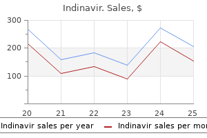
- Levine SR, Welch KM. Cocaine and stroke. Stroke 1988; 19:779.
- King S, Aversano T, Ballard W, et al: ACCF/AHA/SCAI 2007 Update of the Clinical Competence Statement on Cardiac Interventional Procedures, J Am Coll Cardiol 50:82, 2007.
- Modlin IM, Champaneria MC, Chan AK, et al: A three-decade analysis of 3,911 small intestinal neuroendocrine tumors: The rapid pace of no progress. Am J Gastroenterol 102:1464, 2007.
- Yetgin S, Ozen S, Saatci U, et al. Myelodysplastic features in juvenile rheumatoid arthritis. Am J Hematol. 1997; 54:166-9.
- Devine CJ Jr, Horton CE: Chordee without hypospadias, J Urol 110:264n271, 1973.
- Wilson SM: In extremis use a Foley catheter in cardiac stab wound. J Trauma 26:400, 1986.
- Helmstaedter C, Elger CE, Hufnagel A, Zentner J, Schramm J. Different effects of left anterior temporal lobectomy, selective amygdalohippocampectomy, and temporal cortical lesionectomy on verbal learning, memory and recognition. J Epilepsy 9: 39-45, 1996.