

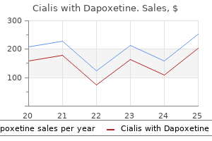
Cialis with Dapoxetine
| Contato
Página Inicial
Steven N. Konstadt, MD, MBa, fa cc
- Chairman
- Department of Anesthesiology
- Maimonides Medical Center
- Brooklyn, New York
- Professor
- Anesthesiology
- Mount Sinai Medical Center
- New York, New York
Conditioning impotence lower back pain buy cialis with dapoxetine now, including flexibility erectile dysfunction medicine list cialis with dapoxetine 40/60 mg purchase visa, strength xalatan erectile dysfunction purchase discount cialis with dapoxetine on-line, and endurance erectile dysfunction jelly order cheap cialis with dapoxetine online, is best performed with a slow impotent rage violet cheap cialis with dapoxetine 20/60mg online, structured interval program. Mazières B, Rouanet S, Guillon Y, et al: Topical ketoprofen patch in the treatment of tendinitis: a randomized, double blind, placebo controlled study. Sellards R, Kuebrich C: the elbow: diagnosis and treatment of common injuries, Prim Care 32:116, 2005. What To Do: Obtain a detailed history of the mechanism of injury, and examine the joint for structural integrity, function, and point tenderness. A tense joint effusion will limit the physical examination (and is one reason to require reevaluation after the swelling has decreased) but also suggests less than a third-degree ligamentous injury, which is normally accompanied by a tear of the joint capsule, and release of any tense effusion. Obtain radiographs (these can be deferred if findings are minimal with full range of motion without bony tenderness or if specific criteria are not met, as for ankle and knee sprains). For first- and second-degree sprains, gently immobilize the joint using an elastic bandage alone or in combination with a cotton roll or plaster splint, as discomfort demands. Dynamic bracing (such as ankle stirrup splints and hinge knee braces) should be used with stable injuries when available. Most upper-extremity injuries can be immobilized by a sling alone or in combination with a soft or rigid splint. Consider prescribing acetaminophen or anti-inflammatory pain medication when the patient complains of pain at rest, and provide crutches when discomfort will not allow weight bearing. Instruct the patient in rest, elevation above the level of the heart, and, when it provides comfort, application of ice 10 to 20 minutes each hour for the first few hours then three or four times a day for 3 days. Explain to the patient that swelling in acute musculoskeletal injuries usually increases for the first 24 hours, and then decreases over the next 2 to 4 days (longer if the treatment above is not employed). Also inform the patient that some swelling and discomfort may persist for several weeks and at times for several months. Advocate early mobilization and early return to normal functions for first- and second-degree sprains. Explain the possibility of occult injuries, the necessity for follow-up, and the slow healing of injured ligaments (usually 6 months until full strength is regained). What Not To Do: Do not obtain radiographs before the history or physical examination. For example, physicians have been steered away from the diagnosis of an avulsion fracture of the base of the fifth metatarsal by the presence of normal ankle films. Discussion A tense joint effusion will limit the physical examination (and is one reason to require reevaluation after the swelling has decreased) but also suggests less than a third-degree ligamentous injury, which is normally accompanied by a tear of the joint capsule, and release of any tense effusion. For this reason, the use of ice should not be mandatory but should be used only when it is comforting to the patient. Suggested Readings Bleakley C, McDonough S, MacAuley D, et al: the use of ice in the treatment of acute soft tissue injury, Am J Sports Med 32:251261, 2004. If comfort allows, gently and repeatedly perform the maneuvers of the McMurray test (see Chapter 115). Obtain knee radiographs, looking for an osteocartilaginous loose body or other disease. With pain and persistent locking, prepare the knee with povidone-iodine solution and, at a point just superior and lateral or medial to the patella, using a 25-gauge, 1-inch needle, inject 10 mL of 0. Leave the patient supine so that gravity will aid in extension and have the patient gently rock and rotate the knee for approximately 20 minutes or until the locked knee has released. Repeated McMurray maneuvers may again be gently performed if joint reduction has not occurred. Alternatively, longitudinal traction can be applied with gentle rotation of the knee internally and externally. When the mechanical block is dislodged and the knee extended, place the patient in a knee immobilizer, keep the patient nonweight-bearing with crutches, and refer the patient to an orthopedic surgeon for early arthroscopic examination and definitive treatment. In such cases, the cause may be torsion of the infrapatellar fat pad or an intraarticular tumor, such as a ganglion. Locking of the knee occurs when one of these structures has become entrapped between the tibial plateau and the femoral condyles, mechanically blocking extension of the joint. If full extension cannot be obtained, the patient can be placed in a soft, bulky, partially immobilizing dressing (thick cotton roll covered with an elastic wrap) and placed on crutches until orthopedic follow-up can be obtained. It is usually better when lying down, worse with movement, and will perhaps radiate around the abdomen or down the thigh but no farther. What To Do: Perform a complete history and physical examination of the abdomen, back, and legs, looking for alternative causes for the back pain. Pay special attention to red flags, such as a history of significant trauma, cancer, weight loss, fever, night sweats, injection drug use, compromised immunity, recumbent night pain, severe and unremitting pain, urinary retention or incontinence, saddle anesthesia, and severe or rapidly progressing neurologic deficit. Red flags on physical examination include elderly patients, fever, spinous point tenderness to percussion, abdominal tenderness or mass, and lower extremity motor weakness. Radiographs are generally not required, but consider obtaining plain radiographs of the lumbosacral spine on patients who have suffered injury that is sufficient to cause bony injury. Mild trauma in patients who are older than 50 years, patients younger than 20 years of age with nontraumatic pain, or patients older than 50 years of age who have had pain for more than a month warrant radiographs. Radiographs should also be ordered for patients who are on longterm corticosteroid medication, patients with a history of osteoporosis or cancer, and patients who are older than 70 years of age. Consider diagnoses such as multiple myeloma, vertebral osteomyelitis, spinal tumor, diskitis, or spinal subdural abscess. Bedside ultrasonography, if the physician is trained in this technique, should be obtained to rule out an abdominal aortic aneurysm. This pain tends to worsen with coughing, Valsalva maneuver, trunk flexion, and prolonged sitting or standing. Look for weakness of ankle or great-toe dorsiflexion (drooping of the big toe and inability to heel walk). Also look for decreased sensation to pinprick over the medial dorsal foot when there is compression of the fifth lumbar nerve root. Look for weak plantar flexion (inability to toe walk), diminished ankle reflex, and paresthesias or decreased sensation to pinprick of the lateral or plantar aspect of the foot when there is first sacral root compression (L5 and S1 radiculopathy account for about 90% to 95% of all lumbar radiculopathies). Raise each leg 30 to 60 degrees of elevation from the horizontal, and consider the test positive for nerve root compression if it produces pain down the leg below the knee along a nerve root distribution, rather than pain in the back. This leg pain is increased by dorsiflexion of the foot and relieved by plantar flexion. Pain generated at less than 30 degrees and greater than 70 degrees is nonspecific. Ipsilateral straight-leg raising is a moderately sensitive but not a specific test. A herniated intervertebral disk is more strongly indicated when contralateral radicular pain is reproduced in one leg by raising the opposite leg. Although controversial, some consultants recommend short-term corticosteroid treatment, such as prednisone, 50 mg qd × 5 days. It should be noted that the 2007 joint guidelines of the American College of Physicians and the American Pain Society recommend against using systemic steroids. The patient should try at least 4 to 6 weeks of conservative treatment before submitting to an operation on the herniated disk. Surgical treatment should be routinely avoided for patients with disk herniation and radiating pain in the absence of neurologic findings. The presence of significant weakness in a myotome is perhaps the most important factor in the decision to perform a relatively early surgical procedure. If the weakness is profound or rapidly progressive, delaying surgery increases the risk for permanent deficit. The rare cauda equina syndrome is the only complication of lumbar disk herniation that calls for emergent surgical referral. It occurs when a massive extrusion of disk nucleus compresses the caudal sac containing lumbar and sacral nerve roots. Bilateral radicular leg pain or weakness, bladder or bowel dysfunction, perineal or perianal anesthesia, decreased rectal sphincter tone in 60% to 80% of cases, and urinary retention in 90% of cases are common findings. For patients who have nonspecific pain that can be treated in an outpatient setting, prescribe a short course of anti-inflammatory analgesics (ibuprofen, naproxen) for patients who do not have any contraindications for using them. A brief course of a muscle relaxant, such as metaxalone (Skelaxin), 800 mg tid to qid (less drowsiness), cyclobenzaprine (Flexeril), 10 mg bid to qid (not recommended for the elderly), or lorazepam (Ativan) 0. The potential benefits must be weighed against the increased rates of dizziness and drowsiness that accompany the use of muscle relaxants. Recommend hot or cold packs (whichever the patient chooses) or alternate both hot and cold. At times sacroiliac dysfunction can cause incapacitating spasms of pain that are precipitated by minor movements or attempts to sit up. The patient will usually be able to localize the pain to the right or left side of the sacrum. When the pain is significant and there are no neurologic findings to suggest nerve root compression or any red flags of underlying systemic disease, it can be quite rewarding to provide an intra-articular injection of a local anesthetic mixed with a corticosteroid. When the needle is in the joint, the needle should advance freely up to its hub without meeting resistance or bony obstruction. There should be a free flow of medication from the syringe without causing soft tissue swelling. If the needle meets any obstruction, reposition it with slight angulation of the needle tip out laterally until the needle advances easily. The patient may feel a brief increase of pain, followed by dramatic relief in 5 to 20 minutes that is usually persistent. Warn the patient that there may be a flare in pain when the anesthetic component wears off that could last for 24 to 48 hours. If the patient gets relief initially, any persistent symptoms should subside over the next 5 to 10 days. This belt may be most helpful during walking and standing activities, but for some patients with significant pain and weakness, wearing it during sedentary activities may also be helpful in reducing symptoms. For point tenderness of the lumbosacral muscles, substantial pain relief may also be obtained by injecting 10 to 20 mL of 0. Quickly puncture the skin, drive the needle in to the muscle belly, and inject the anesthetic, slowly advancing and withdrawing the needle and fanning out the medication in all directions. Often one fan block can reduce symptoms by 95% after injection and yield a 75% permanent reduction of painful spasms. For severe pain that cannot be relieved by injections of local anesthetic, it may be necessary to provide the patient with 1 to 2 days of bed rest, although most patients with acute low back pain recover more rapidly by continuing ordinary activities (within the limits permitted by their pain) than with bed rest or back-mobilizing exercises. Some patients with intractable excruciating pain (especially the elderly) require hospitalization. Refer patients with uncomplicated back pain to their primary care provider for follow-up care in 3 to 7 days. Tell patients that the pain may be recurrent and that cigarette smoking, sedentary activity, and obesity are risk factors for back pain. Teach them to avoid twisting and bending when lifting, and show them how to lift with the back vertical, using thigh muscles and holding heavy objects close to the chest to avoid reinjury. Encourage them to return to work or resume normal activities as soon as possible, with neither bed rest nor exercise in the acute phase, and to participate in an aerobic exercise program when the pain has subsided. Heavy lifting, trunk twisting, and bodily vibration should be avoided in the acute phase. Lumbar supports have not been proven to reduce the incidence of low back pain in industrial workers and should not be routinely recommended for the prevention of low back pain. Do not recommend bed rest for more than 2 days and only recommend it when the pain is severe. Bed rest does not increase the speed of recovery from acute low back pain and sometimes delays recovery. Discussion Low back pain affects men and women equally, with onset most often between the ages of 30 and 50 years. It is the most common and expensive cause of work-related disability in people who are younger than 45 years of age. A definitive diagnosis for nonradiating low back pain cannot be established in 85% of patients because of the weak associations between symptoms, pathologic changes, and imaging results. It can be generally assumed that much of this pain is secondary to musculoligamentous injury, degenerative changes in the spine, or a combination of the two. The approach discussed here is geared only to the management of acute injuries and flare-ups, from which most people recover on their own, which leaves only about 10% developing chronic problems. With acute pain, reassurance plus limited medication may be the most useful intervention. The 90% of back pain patients that become pain free are pain free within 3 months, and more than 90% of those patients recover spontaneously within 4 weeks. Even with diskogenic back pain or disk herniation with radicular pain, there are convincing data to support the nonoperative treatment of these patients in the absence of cauda equine syndrome or progressive neurologic deficit. History and physical examination are essential to rule out serious pathologic conditions that can present as low back pain but require quite different treatment-aortic aneurysm, pyelonephritis, pancreatitis, abdominal tumors, pelvic inflammatory disease, ectopic pregnancy, and retroperitoneal or epidural abscess. Older patients who experience radicular symptoms may have spinal stenosis, which may be accompanied by neurogenic claudication, a syndrome in which pain radiates down the legs, particularly when walking, and is often relieved by rest. This can be distinguished from vascular claudication, because the pain of neurogenic claudication starts even while the patient stands still. The pain is worsened by extension of the spine, which occurs with standing or walking, and improves with flexion, such as sitting or leaning forward. In fact, many radiographic anomalies, such as spina bifida occulta, single-disk narrowing, spondylosis, facet joint abnormalities, and several congenital anomalies, are equally common in symptomatic and asymptomatic individuals. Although adults are more apt to have disk abnormalities, muscle strain, and degenerative changes associated with low back pain, athletically active adolescents are more likely to have posterior element derangements, such as stress fractures of the pars interarticularis. Early recognition of this spondylolysis and treatment by bracing and limitation of activity may prevent nonunion, persistent pain, and disability. Radiation of pain down one or both legs may occur, but usually not below the knee or accompanied by positive straight-leg raising or neurologic deficit. Malingering and drug-seeking are major psychological components to consider in patients who have frequent visits for back pain and whose responses seem overly dramatic or otherwise inappropriate. These patients may move around with little difficulty when they do not know they are being observed.


The posterior urethra causes of erectile dysfunction in 40s cialis with dapoxetine 40/60 mg overnight delivery, approximately 6 cm in length erectile dysfunction kolkata order generic cialis with dapoxetine on-line, is composed of that area which traverses the prostate erectile dysfunction protocol reviews order on line cialis with dapoxetine, which is approximately 34 cm in length impotence questions cialis with dapoxetine 40/60 mg low cost, and that which lies within the confines of the distal sphincter mechanism erectile dysfunction treatment raleigh nc cheap cialis with dapoxetine american express, which is 2 cm in length. At the external border of the distal sphincter mechanism is the junction of the posterior urethra with the anterior urethra. The anterior urethra can be further subdivided in to two areas which are divided on the basis of the areas anterior and posterior to the penoscrotal junction. The bladder neck mechanism lies at the outlet of the bladder around the internal urethral meatus and is composed principally of circularly oriented fibres, although there is a longitudinal component. This sphincter is sufficiently strong to maintain continence even if the distal sphincter mechanism is destroyed. Its principal role, however, is as a genital sphincter causing rapid closure of the bladder neck at the time of emission of semen in to the prostatic urethra. The principal motor control of the bladder neck mechanism appears to be adrenergic via the release of norepinephrine from the sympathetic nerves. Just distal to the bladder neck mechanism is the prostatic urethra, and it must be remembered that the human prostate comprises a significant smooth muscle component. At the apex of the prostate lies the distal sphincter mechanism, which is analogous to the urethral sphincter mechanism of the female and comprises both a lissosphincter and a rhabdosphincter as in the female urethra. Just as for the female sphincter mechanism there is a triple innervation from parasympathetic, sympathetic and somatic nerves (pudendal nerve). In both the male and female urethra a number of glands open in to the posterior urethra and can be the site of infection and source of confusion on occasion at the time of urethrography. There is slight dilatation of the urethra in the bulbar area where the urethra itself is surrounded by the bulbospongiosus muscle. There is a relative constriction of the urethra within the glans penis which helps focus the stream of urine as it comes through the dilatation present at the site of the navicular fossa; this is the narrowest part of the whole urethra. In the prostatic part it is transitional epithelium; in the membraneous and penile parts, pseudostratified and stratified columnar epithelium occur proximally, but, progressing more distally, islands of stratified squamous epithelium appear until in the distal part and the navicular fossa there is a complete sheet of stratified squamous epithelium. Equivalent to the penis in the female is the clitoris, which is also composed of erectile tissue. There are paired seminal vesicles which produce materials, including sugars, needed for sperm to mature and which drain in to the common termination of the vasa deferentia to form the paired common ejaculatory ducts opening in to the prostatic urethra. In addition to the corpus spongiosum which surrounds the urethra, it comprises paired corpora cavernosa which represent the erectile tissue. It must be remembered that at the tip of the penis, the glans penis is contiguous with the corpus spongiosum and abuts against the corpus cavernosum. Penile erection is essential to successful intercourse and is mediated by the nervi erigentes arising from the S2S4 nerve roots. Disorders of both penile and clitoral erection have been increasingly recognised in recent years to be a cause of significant concern within the population, and management of these disorders is an important mainstay of the subspecialty of andrology. There are significant differences between male and female germ cells in terms of timing of production, the number of germ cells produced and their size and shape. In the female, cell division (mitosis) in the stem cells that result in germ cells ceases during embryonic life, and all the oogonia start their first meiotic division before birth. They remain in a resting phase until released from the ovary at ovulation, when the second meiotic division occurs rapidly after the ovum is penetrated by sperm. In contrast, mitosis in the male spermatogonia continues from puberty to old age and death. Cells are always entering meiosis, passing through the two divisions and maturing in to sperm during a process that takes approximately 30 days. In the female, mitosis between oogonia in embryonic life produces a peak population of about six million cells two-thirds of the way through intrauterinal life. There are approximately two million left at birth, which is followed by a dramatic loss of germ cells: by puberty there are only 150 000, and 1,000 are left at the age of 50. In contrast, in the male, large numbers of germ cells persist through life, and in a healthy young man a single ejaculate contains 300 million sperm. The oocyte in the female is one of the largest cells in the body, measuring about 120 m in diameter. In contrast the sperm comprises a head, which is the nucleus, containing tightly packed, condensed genetic material with a small cap. The acrosome and the body contain many mitochondria packed around a central cilium. At the superior (tubal) pole of the ovary is attached a prominent fold of peritoneum, the suspensory ligament of the ovary, which passes upwards over the pelvic brim and external iliac vessels to merge with the peritoneum over psoas major muscle. The ovarian artery gains access to the ovary through the mesovarium and suspensory ligament. A further ligament, the ovarian ligament, runs within the broad ligament to the cornu of the uterus. The upper margin of this fossa is formed by the external iliac vessels, whilst the posterior margin is formed by the ureter and internal iliac vessels. The ovary is very variable in position and may be found prolapsed in to the pouch of Douglas. Relations · · · · structures within the broad ligament; structures on the lateral wall of the pelvis; and abdominal and pelvic viscera. Blood supply the arterial blood supply is from the ovarian artery, which is a branch of the aorta which comes off at the level of the renal arteries. On the left side, it drains in to the left renal vein in a similar fashion to the testicular vein in the male. That part of the cervix lying outside the vagina is related directly to the bladder, whereas the infravaginal cervix has the anterior fornix as an immediate anterior relationship. Posteriorly lies the recto-uterine pouch (of Douglas), which is directly related to the coils of intestine lying in the pouch. Laterally lies the broad ligament; the ureter lies superior and lateral to the supravaginal cervix. The artery then ascends in a tortuous manner, running up the lateral side of the body of the uterus before turning laterally and inferiorly to the uterine tube, where it terminates by anastomosing with the terminal branches of the ovarian artery. Lymphatic drainage Lymphatic drainage of the ovaries follows the ovarian arteries to the para-aortic lymph nodes. Uterus the uterus is a pear-shaped organ which is approximately 7 cm long, 5 cm from side to side at its widest point, and 3 cm anteroposteriorly. The uterine artery also gives off a descending branch which supplies the cervix and the upper vagina. The broad ligament of peritoneum is draped over the fallopian tube like a sheet over a washing line. Each tube comprises the following parts: Lymphatic drainage Lymphatic vessels from the uterus drain in the following manner. Metastases from the fundus of the uterus may, therefore, occur in the inguinal nodes. The cervix drains laterally via the broad ligament to the external iliac nodes, posteriorly in the uterosacral fold to the sacral lymph nodes, and posterolaterally along the uterine vessels to the internal iliac nodes. Its opening is fimbriated and overlies the ovary; ampulla, which is wide, thin walled and tortuous; isthmus, which is narrow straight and thick walled; and intramural part this pierces the uterine wall. Fallopian tubes the fallopian or uterine tubes are 1012 cm long and run from the lateral side of the body of the uterus to the pelvic wall, where they end by opening near the the fallopian tube is covered by peritoneum except for the intramural part. It contains a muscular coat of outer longitudinal and inner circular fibres, and the mucosa is formed of columnar-ciliated cells and lies in longitudinal ridges, each of which is thrown in to numerous folds. This is accomplished by muscular contraction, ciliary action and by the production of a lubricating fluid. The distal end of the tube is open in to the peritoneal cavity, providing direct communication between the peritoneum and the outside and, therefore, a potential pathway for infection. Blood supply the arterial blood supply is from several sources on each side: Broad ligament the broad ligament is a fold of peritoneum which connects the lateral margin of the uterus with the side wall of the pelvis. The broad ligament contains or attaches to the following structures: · · · · vaginal artery; uterine artery; middle rectal artery; and internal pudendal artery, supplying the lower third of the vagina. Venous drainage is via a plexus of veins in the connective tissue around the vagina, draining in to the internal iliac vein. Lymphatic drainage Those from the upper two-thirds of the vagina drain in to the internal and external iliac nodes. In the base of the broad ligament the ureter passes forwards to the bladder lateral to and then immediately above the lateral fornix of the vagina. Each testis is contained by a white fibrous capsule, the tunica albuginea, and is covered by a double serous membrane in to which it became invaginated in fetal life, the tunica vaginalis. Irregular septa arise from the tunica albuginea, dividing the testis in to some 250 lobules, each lobule contains one to three tightly coiled tubules, seminiferous tubules, within which the sperm are produced. The testes lie outside the body because spermatogenesis requires a temperature below that of the body, and if testes fail to descend properly this invariably leads to malfunction in spermatogenesis. At the hilum of the testis the seminiferous tubules drain in to an irregular series of ducts called the rete testis from which efferent tubules arise, transporting the sperm in to the head of the epididymis and subsequently down the epididymis through the vasa, joining with the seminal vesicles prior to forming the common ejaculatory ducts. The epididymis lies along the posterior border of the testis, somewhat to its lateral side. The testis and epididymis each may bear at their upper extremities a small stalked body named respectively the appendix testis (also known as the hydatid of Morgagni) and the appendix epididymis. Vagina the vagina surrounds the cervix of the uterus and then passes downwards and forwards through the pelvic floor to open in to the vestibule, which is the area enclosed by the labia minora and also contains the urethral orifice lying immediately behind the clitoris. The cervix opens in to the anterior wall of the vagina superiorly bulging in to the vaginal lumen. The vagina forms a ring around the cervix, and although this ring is continuous it is divided in to anterior, posterior and lateral fornices. Relations · · · · Anteriorly the cervix enters the vagina above, and below this is the base of the bladder and the urethra which is embedded in the anterior vaginal wall. Posteriorly the posterior fornix is covered by peritoneum in the recto-uterine pouch (of Douglas). Below this the anterior wall of the rectum is immediately posterior to the vagina, and below that the anal canal is separated from it by the perineal body. The venous drainage is by the pampiniform plexus of veins, which becomes a single vessel, the testicular vein, at the deep inguinal ring. Vas deferens the vas deferens (ductus deferens) commences at the inferior pole of the testis as the continuation of the epididymis. It is a thick muscular tube which transports sperm from the epididymis to the ejaculatory ducts within the prostate gland. It passes through the scrotum, inguinal canal and comes to lie on the side wall of the pelvis. The vas ends by uniting with the ducts of the seminal vesicle to become the common ejaculatory duct. Lymphatic drainage Lymphatic vessels from the testis and epididymis accompany the testicular veins to drain in to the para-aortic nodes. The following structures are encountered: Prostate the human prostate surrounds the prostatic urethra. There are two principal components to the prostate: the glandular component and a smooth muscle component. Approximately 25% of normal prostate is composed of smooth muscle, and with development of benign enlargement of the prostate, contrary to popular perception, although this is a glandular hyperplasia there is also a relative increase in the amount of smooth muscle such that smooth muscle in the hyperplastic prostate constitutes 40% of the gland. The majority of the prostate lies on the lateral and posterior aspects of the urethra, with little anterior prostatic tissue. Once the parietal layer of the tunica vaginalis has been divided, the visceral layer of the tunica vaginalis is seen covering the white tunica albuginea. Skin Dartos muscle Relations External spermatic Cremasteric fascia Internal spermatic fascia Tunica vaginalis (parietal layer) Tunica vaginalis (visceral layer) Vas deferens · · · · · · Anteriorly the pubic symphysis is separated by the extraperitoneal fat of the retropubic space. Inferiorly the apex of the prostate rests on the external urethral sphincter within the deep perineal pouch. Clinically the prostate gland is divided in to lobes: the posterior lobe lies posterior to the urethra and inferior to the plane defined by the course of the ejaculatory ducts. Two lateral lobes (or right and left lobes) are separated by a shallow posterior median groove which can be felt on rectal examination. Blood supply the arterial blood supply is derived from the inferior vesical artery, which is a branch of the internal iliac artery. The venous drainage is via the prostatic venous plexus, which drains in to the internal iliac vein on each side. Some blood drains posteriorly around the rectum to the valveless vertebral veins of Batson. This is said to explain why prostatic carcinoma metastasises early to the bones of the lumbar spine and pelvis. The prostatic urethra is the widest portion of the urethra, and on its posterior wall there is a prominent bulge known as the urethral crest, at the distal end of which lies the verumontanum, which has a midline opening on it leading to the prostatic utricle or uterus masculinus, which arises from the Mullerian ducts. Lying on either side of the opening of the utricle is the termination of the ejaculatory ducts, where seminal emission occurs. The prostate has an important physiological role in producing secretions which are important to the survival and function of spermatozoa. It should not be forgotten that prostaglandins were so named having been first identified in the prostate. Each seminal vesicle has a common drainage with its neighbouring vas via the common ejaculatory duct. A subsidiary but extremely important role is that of the production of certain hormones. Approximately one-fifth of the cardiac output passes through the kidneys each minute, resulting in a renal blood flow of up to 400 mL per 100 g of kidney per min (650 mL/min per kidney). The renal blood pressure remains extremely constant despite profound changes in systemic blood pressure, and the survival advantage of this mechanism is apparent on reflection. This phenomenon is described as autoregulation and is principally mediated via effects on preglomerular vascular resistance. Whilst the underlying mechanism is the subject of intensive study, it is thought to be related to intrinsic myogenic tone within blood vessels, independent of neural factors.

It is an organochloride compound that is absorbed and can be toxic to the central nervous system and cause anemia drinking causes erectile dysfunction purchase cialis with dapoxetine 20/60 mg amex. In view of reports of lindane resistance and the availability of products with more favorable safety profiles erectile dysfunction at the age of 25 best order for cialis with dapoxetine, it is no longer recommended generic erectile dysfunction drugs online order 20/60 mg cialis with dapoxetine free shipping. Discussion Blood-sucking lice have long been successful obligate parasites of humans erectile dysfunction viagra cialis levitra cialis with dapoxetine 40/60mg buy low price. Patients with louse infestation present with pruritus erectile dysfunction doctors in louisville ky buy 40/60mg cialis with dapoxetine fast delivery, excoriations, and lymphadenopathy. After attaching to the patient, the louse inserts its mouth parts and injects saliva with vasodilatory properties. An inflammatory reaction to injected louse saliva has been suggested as the most likely cause of bite reactions. Transmission in most cases occurs by direct contact with the head of another infested individual. Except in very humid climates, lice lay nits (ova within a chitinous case) within 1 to 2 mm of the scalp. Young lice hatch within 1 week and pass through three nymphal stages, maturing to adults over a period of 1 week. In most climates, they survive only several hours off the scalp, although they may live for up to 4 days in favorable conditions. In the United States, blacks have a lower incidence of infestation, possibly because lice are better adapted to grasp the more cylindrical hairs of whites or Asians. The diagnosis of head lice is definitive when crawling lice are seen in the scalp hair or are combed from the scalp. Because head lice avoid light and can crawl quickly, the use of louse combs increases the chances of finding live lice. Nits alone are not diagnostic of active infestation, but if the nits are within 1 cm (¼ inch) of the scalp, active infestation is likely. A parent, teacher, or school nurse generally notices them and mistakes them for nits. Among presumed "lice" and "nits" submitted by physicians, nurses, teachers, and parents to a laboratory for identification, many were found to be artifacts such as dandruff, hairspray droplets, scabs, dirt, or other insects. If newly hatched eggs that survived the initial therapy are not re-treated, the cycle may repeat itself every 3 weeks. Although there is no proven transmission from fomites, such as brushes, hats, combs, linens, and stuffed animals, head lice and ova have been found on such items; therefore it is probably expedient to eradicate these lice by vacuuming, washing, dry cleaning, or isolating items in sealed plastic bags for 2 weeks. No healthy child should be excluded from or allowed to miss school time because of head lice. Pubic lice are distinct in appearance from head and body lice; they have short crablike bodies. A major concern in treating crab lice is the lack of appreciation for their tendency to reside in rectal hairs. Some dermatologists prefer to treat pubic lice using ivermectin as their first-line therapy. Besides sexual transmission, pubic lice may also be acquired by sharing a bed with an infested person. Children with pubic lice have usually been infected through contact with an adult. In the United States, the body louse has become less common in the general population. Body lice infestation in all developed countries is generally seen among the homeless in urban areas. It also is common among refugees and those who live in crowded conditions or cannot launder their clothing. Worldwide, body lice are important vectors for louse-borne relapsing fever, trench fever, and epidemic typhus, especially among refugees. The body louse and nits are generally found in the clothing seams of a parasitized individual, but the louse grabs on to body hairs to feed. There is no change for a period of several days to a few weeks; then the generalized rash appears, composed of crops of small (0. The condition may be asymptomatic or accompanied by varying degrees of pruritus (25% of patients have mild to severe itching). The lesions will gradually extend in size and may become confluent with one another. Transient worsening of the rash or a second wave of lesions is not uncommon until eventual spontaneous resolution of the eruption. Pityriasis rosea can have a distinctly different appearance on patients with brown skin or dark skin. The herald patch, as well as the diffuse rash that follows, may have a gray, dark brown, or even black appearance. There may be either hypopigmented or hyperpigmented areas visible after the lesions resolve. Be sympathetic, and let them know that it is understandable how frightening it can seem. In addition, inform them about the need to contact their physician if the rash or pruritus lasts more than 3 months. Acyclovir may be effective in the treatment of pityriasis rosea, especially in patients treated within the first week of onset of the generalized rash. At the moment, however, no treatment can be recommended on the basis of evidence-based medicine. If the diagnosis is uncertain, especially if the palms and soles are affected and the patient is sexually active, draw blood for serologic testing for syphilis. What Not To Do: Do not have a biopsy performed when findings are typical for pityriasis rosea. These are effective only in the most severe inflammatory varieties of this syndrome. Do not send off a serologic test for syphilis without ensuring that the results will be seen and acted on. It has been described in the medical literature for more than 200 years but was given its current name by Camille Gilbert in 1860. Viral and bacterial causes have been sought, but convincing answers have not yet been found. Results of a controlled trial (neither randomized nor double-blind) with oral acyclovir suggest that early treatment with this antiviral is justified, because it apparently reduces the duration and severity of symptoms. Malaise, nausea, loss of appetite, headache, difficulty in concentration, irritability, gastrointestinal and upper respiratory symptoms (up to 69%), joint pain, swelling of lymph nodes, sore throat, and mild fever are often, although inconsistently, reported. The "herald patch" may not be seen in 20% to 30% of cases, and there are many variations from the classic presentation described. Infrequently, oral lesions will accompany the skin rash and resolve along with it: these include punctate hemorrhages, erosions, ulcerations, erythematous macules, annular lesions, and plaques. Other diagnostic considerations besides syphilis include tinea corporis, seborrheic dermatitis, guttate psoriasis, and tinea versicolor. Drago F, Vecchio F, Rebora A: Use of high-dose acyclovir in pityriasis rosea, J Am Acad Dermatol 54:8285, 2006. Drago F, Broccolo F, Alfredo Rebora A: Pityriasis rosea: an update with a critical appraisal of its possible herpesviral etiology, J Am Acad Dermatol 61:303318, 2009. Jones D: the young adult: common inflammatory skin disorders, Clin Fam Pract 5:627652, 2003. The head, neck, and extremities are most commonly involved, especially the lips, oral mucosa, and fingers. An unsightly lesion forms, beginning as an extremely friable red or yellow papule or polyp, which now bleeds with every slight trauma. Objective findings usually include a crusted, sometimes purulent-appearing collection of erythematous, well-demarcated, red granulation tissue arising from a moist, sometimes hemorrhagic, skin ulceration, often with a collarette of scale at the base. What To Do: Cleanse the area with an agent such as hydrogen peroxide or povidone-iodine solution. The base of the lesion can be injected with 1% lidocaine with epinephrine to minimize the resulting hemorrhage. Thermal cautery may also be used, but local anesthesia will probably be required because of the potential inadvertent contact with intact dermis. For pedunculated lesions, an atraumatic, simple, fast, and cost-effective alternative that does not require anesthesia is to ligate the base of the granuloma using a soft, absorbable, surgical suture material. This can be covered by a simple wound dressing, and the tumor can be expected to become necrotic and fall off in several days. Inform patients or parents that although uncommon, bleeding could occur, and simple continuous compression for several minutes will control any minor hemorrhage. If the ligature does not reach far enough down to include the nurturing vessel, part of the granuloma may persist. This smaller lesion can then be easily treated with silver nitrate or thermal cautery. Patients with large lesions can be referred for treatment with a pulsed-dye laser. Warn the patient about the potential signs of developing infection and the need to return for treatment. Patients and parents should also be alerted to the possibility of recurrence after removal. Dermatology referral is recommended if the lesion recurs or multiple satellite lesions occur after excision. What Not To Do: Do not cauterize or ligate any lesion that by history and appearance might be neoplastic in nature. Pyogenic granuloma is occasionally confused with amelanotic melanoma, basal cell carcinoma, and squamous cell carcinoma. Suspicious lesions should be referred for complete excision and pathologic examination. Discussion Pyogenic granulomas, also known as lobular hemangiomas, are common vascular tumors of the skin and mucous membranes usually seen in children and young adults. Their cause is unknown, but pyogenic granuloma is a misnomer, because it is probably not infectious (pyogenic) in origin. It has been argued to be inflammatory and hyperplastic rather than a true neoplasm. Rapid growth is in response to an unknown stimulus that triggers endothelial proliferation and angiogenesis. They can be solitary or multiple and can arise within preexisting lesions, such as spider angioma and port wine stain. Although a minority of pyogenic granulomas involute spontaneously within 6 months, most patients seek treatment because of bleeding. Gingival lesions are common in pregnant women, in whom these lesions are called epulis gravidarum. It is not uncommon for secondary cellulitis to develop after the pyogenic granuloma is cauterized. It is therefore reasonable to place a patient on a short course (3 to 4 days) of a high-dose antibiotic (dicloxacillin or cephalexin, 500 mg qid, or cefadroxil, 500 mg bid) when the wound is located on a distal extremity. Infants and young children may also have palm and sole involvement with vesicular and pustular lesions. Scabies in the elderly may be difficult to diagnose because the cutaneous lesions are often very subtle. What To Do: Attempt to confirm the diagnosis of scabies by placing mineral oil over five or six nonexcoriated suspicious papules or burrows, and scrape or shave them with a No. Examine under low magnification for the mite, its oval eggs, or fecal concretions (scybala). If the clinical picture alone is convincing, especially if there is someone else at home (a source) who is itching, treatment should be instituted without the help of microscopic examination, or even in the face of negative scrapings. Prescribe prescription-strength permethrin (Elimite) 5% insecticidal cream, 60 g, for the patient to massage from his head to the soles of his feet at bedtime, and have him leave it on for 8 to 14 hours before washing it off the next morning. The patient should trim fingernails, scrub beneath them, and then apply the scabicide under the nails. A total of two or more doses at least 7 days apart may be necessary to eliminate a scabies infestation. The safety of ivermectin in children weighing less than 15 kg and in pregnant women has not been established. The high cost and extensive surface area of application that is necessary lead to poor compliance, especially in elderly patients. Massage cream/lotion in to entire body from chin down, repeat 24 hours later and then bathe 48 hours later. Sulfur is the oldest known treatment of scabies, and it is the drug of choice for infants younger than 2 months of age and for pregnant or lactating women. The cream is applied nightly for three consecutive nights and washed off 24 hours later. The major drawback to sulfur treatment is the unpleasant odor; also, the treatment will often stain clothes. Secondary infection from scratching, such as impetiginized excoriations, can be treated with mupirocin (Bactroban) cream 2%. Folliculitis, abscess formation, lymphangitis, and cellulitis should be treated with appropriate drainage and antibiotics (see Chapters 163, 166, and 172). Tell the patient that the itching will not go away immediately and that this does not mean that the treatment was ineffective. Dead mites and eggs continue to cause an immune response but will eventually be eliminated during normal cutaneous turnover. An antipruritic agent, such as hydroxyzine (Atarax, Vistaril), 25 to 50 mg q6h, can be prescribed for comfort. Adding a short course of oral prednisone may be most effective when pruritus is severe. Clothing, bedding, and towels should be washed with hot water or dry cleaned or placed through the heat cycle of a dryer to prevent reinfection.

This results in a degree of plasticity which is relevant to malignant infiltration whereby uni-directional lymph flow may be diverted due to blockage at proximal sites by tumour emboli impotence treatment discount cialis with dapoxetine 20/60 mg buy online. The lymphatics of the skin of the breast represent part of the superficial system of the neck erectile dysfunction adderall generic cialis with dapoxetine 40/60 mg buy, thorax and abdomen erectile dysfunction medicine in bangladesh generic cialis with dapoxetine 40/60mg overnight delivery. From this subareolar and a related circumareolar plexus impotence divorce cialis with dapoxetine 40/60 mg order line, lymph flows principally to the axillary nodes via a lateral lymphatic trunk impotence over 50 purchase cialis with dapoxetine 20/60 mg free shipping. This together with the minor inferior and medial lymphatic trunks drain along the surface of the breast to penetrate the cribriform fascia and reach the various groups of axillary nodes. Lymph drains medially from the circumareolar plexus in to lymphatics accompanying the internal mammary vessels to enter the internal mammary (parasternal) nodes. Approximately 75% of lymph flow passes to the axillary nodes and 25% to the internal mammary nodes. Patients now present with smaller tumours where there is less likelihood of nodal involvement (2530%). Sentinel lymph node biopsy is a new staging procedure which can accurately determine the pathological status of axillary nodes. Clearance of nodal tissue is confined to clinically node positive patients and those clinically node negative patients found to have nodal disease on sentinel node biopsy. Some of these directly influence physiological processes whilst others act in a permissive capacity, but collectively they are important for Lymphatic vessels the lymphatic system of the breast is a complex network of arborising vessels which reflect its embryological origin from anterior thoracic wall structures. A the lymphatics of the nipple are large and numerous and drain along the surface of the breast beneath the skin. B the deeper lymphatics of the breast interconnect and drain chiefly in to main channels directed towards the axilla. Oestrogen promotes duct elongation and branching as well as fat deposition; progesterone governs lobule development and establishes the secretory state. During pregnancy, the alveoli further develop under the influence of prolactin which triggers milk production and release after parturition. Precursor tissue resembling sweat glands passes through a branching stage to form 1525 epithelial strips which later canalize to form ducts which ultimately differentiate to the end-vesicle stage of the newborn infant. This primitive duct system starts peripherally and grows towards the centre with 610 ducts converging upon the nipple at birth. Maternal progesterone stimulates lobular development with end-vesicle formation but there is much variation in the degree of morphological differentiation in the neonate with some having a secretory epithelial phenotype. After birth, the abrupt withdrawal of female sex hormones allows endogenous prolactin to act directly on the alveolar epithelium. There may be a transient hyperplasia of the breast epithelium in the immediate post-natal period, but subsequently the breast tissue regresses and involutes with loss of end-vesicles. During childhood there is only minimal branching of the ducts with no alveolar formation. These gonadotrophins are released from the pituitary in response to hypothalamic gonadotrophin-releasing hormone. Breast epithelium is initially stimulated by oestrogen only (oestradiol) which results in ductal growth and branching. Defined histological changes occur within breast tissue which correlate with stages of the cycle; during the proliferative phase, oestrogens are secreted by the maturing Graafian follicle and stimulate epithelial mitoses with proliferation and budding of ducts. Concomitant connective tissue hyperplasia and progesterone secretion in the luteal phase results in increased blood flow, lobular oedema and thickening of the basement membrane. This together with enlargement of alveolar diameter and appearance of secretions in response to elevated prolactin causes a cyclical increase in breast size of 1530 mls. At the end of each menstrual cycle, regressive changes occur with shrinkage of the lobulo-alveolar units and narrowing of lumina. Progesterone also inhibits ductular growth and stimulates mesenchymal elements with a massive increase in blood supply. During the third trimester this increased vascularity coupled with distension of colostrum-laden alveoli leads to an increase in breast size and an average weight gain for each breast of 350 g. It is a complex process under the hormonal control of the hypothalamus, anterior pituitary, ovary, placenta and adrenals. Prolactin, or lactogen is the most powerful hormone responsible for milk production, but the secondary actions of cortisol, insulin and growth hormone are permissive. Though there is an increase in the protein synthesizing capacity of alveolar epithelial cells with activation of pathways for protein, milk fat and lactose production, minimal secretion occurs in to the alveolar lumina. High circulating levels of oestrogen and progesterone during pregnancy suppress development of prolactin receptors on alveolar cells and thus prevent manifestation of the full secretory pheno-type until after parturition. Secretion of mature milk begins approximately 3040 hours after birth and although prolactin levels return to near normal, suckling induces periodic surges of prolactin release from the anterior pituitary via a neuro-endocrine reflex involving signals relayed from sensory nerve endings in the breast to the hypothalamus. Suckling can maintain lactation indefinitely and averages 12 mls/g of breast tissue per day. During the first few days post-partum the immature milk product colostrum is secreted. This is a thick yellow substance which differs from mature milk in containing a high protein content in the form of lactoglobulin. IgG is secreted in to the colostrum which thus provides passive immunity in addition to nutrition for the newborn infant. This occurs in two distinct phases and is driven by a large surge of sex steroids from the fetoplacental unit (human placental lactogen and human chorionic gonadotrophin) together with prolactin and growth hormone from the anterior pituitary gland. Phase I development In the early stages of pregnancy there is proliferation of lobulo-alveolar epithelium with a marked increase in the number of alveoli per lobule (up to ten-fold). More lobules are formed and there is ductal sprouting and elongation which far exceeds that seen in the menstrual cycle. A more differentiated form of lobule characterizes the end of the first trimester when there is noticeable breast enlargement and a sensation of breast heaviness and fullness. There is often venous engorgement in the overlying skin and the nipple and areola become more pigmented and the areola enlarges. Milk letdown Stored milk cannot be released by suction alone; the act of suckling involves tactile stimulation of the nipple with transmission of impulses to the hypothalamus via spinal reflexes (T4, T5 and T6 sensory afferent nerve roots). Myoepithelial cells surrounding acini contract with expulsion of milk in to the lactiferous sinuses. Oxytocin also causes contraction of smooth muscle around these major ducts which encourages expression of milk. Secretion of oxytocin can be modulated by physiological factors; anticipation of breast feeding increases it whilst pain and anxiety inhibit secretion. When suckling stops, prolactin levels are no longer stimulated and the breast involutes to its pre-partum state. Accumulation of milk and lobulo-aveolar distortion is thought to trigger this process which leads to a decrease in size of lobules. The increased fat and connective tissue within the breast often remain, so that the breasts remain larger. Sometimes, however, there is atrophy of all elements of the breast, resulting in shrinkage to a size smaller than before. Within the remaining lobules there is thinning of epithelial and myoepithelial layers, thickening of the basal lamina and obliteration of the lumina. This process may culminate in a scattering of atrophic ducts and acini within a hyalinised stroma. The epithelium of the terminal ducts rather than the acini is considered more prone to neoplastic transformation and more susceptible to environmental factors which initiate malignant change. Lobules are embedded in a loose specialized connective tissue stroma which is itself altered in certain disease states. This reflects poor understanding of certain conditions and clinical overlap in patterns of symptoms resulting from physiological changes and abnormal or disease states. In order to be useful, any system of description should incorporate both clinical and histopathological features. Many of the breast complaints of pain, lumpiness (nodularity) and nipple discharge may be sequelae of normal physiological changes taking place at different stages of reproductive life and in response to cyclical hormonal changes. This scenario is further compounded by similarities in symptoms and signs of benign and malignant pathological conditions which can be clinically indistinguishable. This is the basis of triple assessment in which clinical evaluation is complemented by radiological imaging and some form of tissue biopsy. Benign non-neoplastic conditions of the breast display a wide variety of proliferative and regressive changes within breast tissue. Many cases of breast pain and lumpiness can be attributed to fibrocystic change which might be referred to as fibrocystic disease when symptoms are severe. It is often perceived as a non-specific condition provided discrete lump such as cancer, fibroadenoma or a cyst has been excluded. Cyclical pain and nodularity is very common in women of reproductive age and is a disturbed response to the hormonal environment of the breast. Symptoms are greatest in the pre-menstrual phase when levels of oestrogen and progesterone are highest. This creates a walled space filled with fluid which can vary in size from a few millimeters (microcyst) to several centimeters (macrocyst). Lining epithelial cells tend to be large with abundant granular eosinophilic cytoplasm and basal nuclei (apocrine metaplasia). Diagnosis is confirmed by aspiration and cytology is indicated only if the aspirate is blood-stained (intracystic papilloma or carcinoma) or the cyst refills. Cysts constitute up to 15% of all discrete breast lumps and do not predispose to cancer; adenosis an increase in the number of acini or ductules within a lobule without thickening of the ductular epithelium. It is usually of the blunt duct type in which alveoli showed marked dilatation and an irregular outline; sclerosing adenosis this is an abnormality of stromal involution leading to localized proliferation of both stroma and acini. There is prominent mitotic activity but no dysplasia and the lobules are distorted with infiltrative margins. Hyperplasia is an increase in the number of cell layers above the basement membrane. In mild forms, spaces are lined by 34 cell layers, whilst in moderate to severe forms this exceeds 4 cells in thickness and there may be proliferating cell masses distending and distorting involved spaces. Atypical hyperplasia refers to lesions with both an overgrowth of epithelium and cytological atypia. These two pathological entities are often grouped together as lobular neoplasia as they both distend the acini within a lobular unit to varying extent. The thickness of the ductular epithelium is much greater than normal, and the cells exhibit malignant characteristics: namely, variations in size and shape of cells and nuclei and a broad range of nuclear staining. Mitoses are also evident, but there is no invasion of the basement membrane in this section. Fat necrosis Atypical hyperplasia confers an absolute risk of invasive breast cancer of 810% at 1015 years. Subsequent inflammation and fibroelastic reactions can produce a hard irregular lump tethered to skin which mimics a carcinoma. Periductal inflammation occurs in perimenopausal women and is often sterile, at least initially. Puerperal breast abscesses occur during or soon after lactation and are usually pyogenic with the causative organism being Staphylococcus aureus. Infection probably is introduced via cracked or traumatized nipples during suckling. Infection commences within the main lactiferous ducts producing local inflammation which progresses to a generalized cellulitis affecting one radial section of the breast. At this stage, when there is no focal collection of pus, the infection can be successfully treated with intravenous anti-staphylococcal agents. However, once abscess formation occurs, pus must be surgically drained in order for resolution of the inflammatory process. If surgical intervention is deferred, then the combination of inflammation and scarring can destroy a large part of the breast parenchyma. They arise from a single lobule rather than a single cell and respond to cyclical hormonal changes within the breast. Most undergo spontaneous regression; small fibroadenomas can be subclinical and discovered incidentally on imaging (commonest cause of a breast lump under 30 years of age). Duct ectasia this is an involutional change characterized by dilatation and shortening of the subareolar ducts. More complex abscesses should be drained via an incision placed some distance from the areolar and the wound closed around a corrugated drain which is left in situ for a few days. Perhaps ironically, the management of in situ carcinoma is more controversial than its invasive counterpart and may necessitate mastectomy. However, enlargement is predominantly due to an increase in stromal tissue and should be distinguished from pseudogynaecomastia caused by deposition of subcutaneous fat. The condition is unilateral or bilateral and most cases are idiopathic with no identifiable cause. Physiological gynaecomastia is seen in the newborn (under the influence of maternal hormones) and at puberty when there is a temporary increase in the oestrogen/androgen ratio. This type of gynaecomastia resolves spontaneously, though the pubescent type can be a source of embarrassment. Pathological causes of gynaecomastia include liver disease, lung disease (bronchiectasis, chronic obstructive airways disease, tuberculosis) and testicular tumours. As the process involves mainly the ductules of the lobules, the term ductal carcinoma is used, but this refers to a histological pattern and not tissue of origin. Ductal carcinoma in situ this is a complex disease entity with several histological variants, including comedo, cribriform, solid and micropapillary. Dystrophic deposits of calcium produce coarse linear branching calcification on mammography. Neoplastic cells lining the ducts are usually arranged as solid sheets with central necrosis.
Order 40/60mg cialis with dapoxetine otc. Dr. Sebi speaks on fasting and its ability to cure impotence.
References
- McAllister DA, Maclay JD, Mills NL, et al. Arterial stiffness is independently associated with emphysema severity in patients with chronic obstructive pulmonary disease. Am J Respir Crit Care Med 2007; 176: 1208-1214.
- Hoffman MS, De Cesare SL, Roberts WS, et al. Upper vaginectomy for in situ and occult, superficially invasive carcinoma of the vagina. Am J Obstet Gynecol 1992;166(1 Pt 1):30-33.
- Rakha E, Puls F, Saidul I, et al: Torsion of the testicular appendix: importance of associated acute inflammation, J Clin Pathol 59:831n834, 2006.
- Nicolaides K, Brizot M de L, Patel F, Snijders R. Comparison of chorionic villus sampling and amniocentesis for fetal karyotyping at 10-13 weeks gestation. Lancet 1994; 344: 435-9.